9.5 Alzheimer’s Disease
Open Resources for Nursing (Open RN)
Dementia is a general term that refers to a decline in cognitive ability severe enough to interfere with a person’s ability to complete their activities of daily living. Alzheimer’s disease (AD) is the most common type of dementia, accounting for at least two thirds of cases of dementia in people aged 65 and older. AD is a neurodegenerative disease that causes progressive and disabling impairment of cognitive functions, including memory, comprehension, language, attention, reasoning, and judgment. An estimated 6.7 million Americans are living with AD.[1]
Family members commonly function as unpaid caregivers to support individuals with AD who continue to live at home, including assistance with activities of daily living (ADLs), paying bills, shopping, and preparing meals. Caregivers also provide emotional support, help manage health conditions, and ensure safety at home. Caregiving is time-intensive and can be physically and emotionally exhausting, causing declines in the caregiver’s health.[2] Nurses often address caregiver health when caring for clients with AD.
Pathophysiology
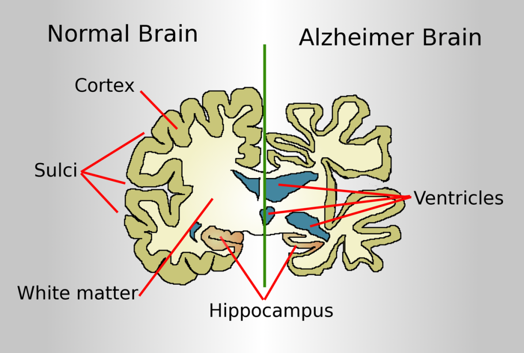
The brain of an older adult experiences normal physiological changes associated with aging, such as increased size of the ventricles. However, with Alzheimer’s disease and other forms of dementia, these changes occur at a much more rapid pace, along with significant atrophy of the cerebral cortex and degeneration of neurons. See Figures 9.12[3] and 9.13[4] for illustrations comparing a normal brain to a brain of a person with Alzheimer’s disease.

The hallmark microscopic changes of AD consist of neurofibrillary tangles and neuritic plaques. Neurofibrillary tangles consist of fibrous tissue that impair the transmission of impulses from neuron to neuron. Neuronal transmission is further hindered by neuritic plaques, composed of abnormal proteins called beta amyloid and tau, that damage the neurons. See Figure 9.14[5] for an illustration of neurodegeneration caused by these abnormal proteins.
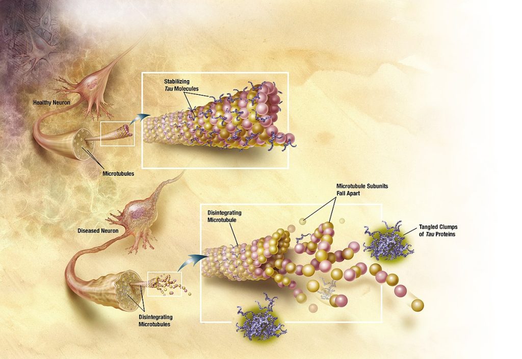
Initial neuron damage occurs in the parts of the brain responsible for memory, language, and thinking, so initial symptoms of Alzheimer’s disease are short-term memory loss and language and thinking problems. Brain changes that cause these symptoms are thought to begin 20 years or more before symptoms start.[6]
Imbalanced neurotransmitter levels are also associated with Alzheimer’s disease. For example, decreased levels of acetylcholine (ACh), norepinephrine, dopamine, and serotonin can further contribute to impaired cognition, loss of short-term/recent memory, and ability to retain new memories.
Risk Factors
The cause of Alzheimer’s disease is not certain. However, age, gender, and genetics are three major risk factors linked to AD, with increasing age being the greatest risk factor. The percentage of people with AD increases dramatically with age, with 33% of people aged 85 or older having AD. Females over 65 years old are more likely to develop AD than men, and having a first-degree relative with AD also increases one’s risk. There is also an increased incidence of AD in people who have Down’s syndrome. Certain medications are associated with higher risk for AD, including benzodiazepines, anticholinergics, and long-term use of proton pump inhibitors.[7],[8] Other risk factors may also play a role, such as exposures to toxic chemicals and immunological changes.[9],[10]
Although age, gender, and genetics cannot be changed, some risk factors can be modified to reduce the risk of cognitive decline and dementia. Examples of modifiable risk factors are participating in physical activity, stopping smoking, staying socially and mentally active, maintaining blood pressure in a normal range, and following a healthy diet.[11]
Some research also indicates that additional years of formal education builds cognitive reserve. Cognitive reserve refers to the brain’s ability to make flexible and efficient use of networks of neuron-to-neuron connections that enable a person to continue to carry out cognitive tasks despite degenerative brain changes. In addition to years of formal education, having a mentally stimulating job and engaging in other mentally stimulating activities may also help build cognitive reserve.[12]
Research on the impact of ethnicity finds a lower incidence of dementia in African Americans, but those diagnosed with dementia showed greater cognitive deficits, neuropsychiatric symptoms, severity of symptoms, and functional dependence.[13]
Evidence indicates that even mild traumatic brain injury (TBI) increases the risk of developing certain forms of dementia. A recent meta-analysis estimates that any form of TBI increases the risk for dementia by nearly 70%. Post-traumatic stress disorder (PTSD) also increases the risk of dementia and is two to five times more common in veterans compared with the general population.[14]
A recent nationwide research study called New IDEAS (Imaging Dementia—Evidence for Amyloid Scanning) is looking for better ways to diagnose and care for people from diverse backgrounds who are experiencing changes in their memory, including mild cognitive impairment and dementia.[15] The goal of the New IDEAS study is to ensure that its results represent all racial and ethnic groups. As new treatments are being developed, the research hopes to provide information for safe and effective care for everyone, regardless of race, ethnicity, age, or gender.[16]
Read additional information in the 2023 Alzheimer’s Disease Facts and Figures PDF by the Alzheimer’s Association.
Read about the Alzheimer’s Association’s New IDEAS Study.
Stages of Alzheimer’s Disease
The initial presenting symptom of AD is impaired short-term memory although long-term memory remains intact. Impaired short-term memory is followed by impaired problem-solving, judgment, executive functioning, motivation, and organization, leading to problems with multitasking and abstract thinking.
Alzheimer’s disease is classified into these different states based on the degree of cognitive impairment: mild, moderate, and severe[17]:
- Mild: Most people with mild AD are able to function independently but require assistance with some activities to maximize their independence and remain safe. Handling finances and paying bills may be especially challenging, and they may need more time to complete common daily tasks. They may still be able to drive, work, and participate in their favorite activities.
- Moderate: Individuals with moderate AD experience additional problems with memory and language, are more likely to become confused, and find it harder to complete multistep tasks such as bathing and dressing. They may wander, become incontinent, and have difficulty sleeping. Personality and behavioral changes may occur, including suspiciousness, agitation, and depression. They may experience increased confusion at night or when lighting is inadequate, referred to as sundowning. They may also begin to have difficulty recognizing loved ones.
- Severe: The ability to verbally communicate becomes greatly diminished in people with severe AD, and individuals are likely to require around-the-clock care. Because areas of the brain involved in movement become damaged, individuals become bed bound. Being bed-bound makes them vulnerable to additional medical complications resulting from immobility, including blood clots, pressure injuries, skin infections, and sepsis. Damage to areas of the brain that control swallowing causes dysphagia and can result in aspiration pneumonia, a contributing cause of death among many individuals with AD.
The symptoms of Alzheimer’s disease worsen over time. On average a person with Alzheimer’s disease lives four to eight years after diagnosis, but depending on variable factors, they may live as long as 20 years.
Assessment
Obtaining a thorough health history and physical assessment is important to distinguish AD from other forms of dementia and acute confusion, referred to as delirium. Interviewing family members or significant others may be necessary because clients with dementia may be unaware of their symptoms. Assessment should include the onset, duration, progression, and course of symptoms, as well as a complete functional status assessment.
Neuropsychological Tests
Neurologists or psychologists may administer several types of neuropsychological tests to determine the extent of the impaired cognition and to measure changes over time. The type of test used is based on the ability of the client.[18]
A common neuropsychological test is the Mini Mental State Exam (MMSE). The MMSE assesses five major cognitive areas, including orientation, registration, attention and calculation, recall, and speech-language-reading.
The MMSE test requires the ability to read. For clients who are unable to read, a test called the set test may be used. In the set test, the client is asked to name ten items in each of four sets: fruits, animals, colors, and twins. The client receives one point for each item out of 40 possible points. A score above 25 indicates the client does not have dementia.
View an example MMSE test: The Mini Mental State Examination (MMSE) PDF.
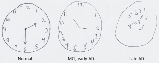
Another cognitive screening tool that does not require the ability to read is the clock drawing test. The client is provided with a piece of paper with a pre-drawn circle approximately 10 cm in diameter and told that the circle represents the face of a clock. The person is asked to write numbers within the circle so that it looks like a clock and then add arms to the clock to show a desired time, such as “ten minutes after eleven.” See Figure 9.15[19] for an illustration of the clock drawing test with results from clients with different states of AD.
Laboratory and Diagnostic Tests
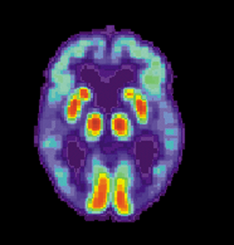
A definitive diagnosis of Alzheimer’s disease can only be made by autopsy and examination of brain tissue showing the presence of neurofibrillary tangles and neuritic plaques. However, recent progress has been made in measuring these brain changes with diagnostic tests. For example, abnormal levels of beta-amyloid and tau can be measured in cerebrospinal fluid (CSF), and positron emission tomography (PET) can produce images showing where beta-amyloid and tau have accumulated in the brain.[20],[21] See Figure 9.16[22] for an image of a PET scan of a person with Alzheimer’s disease showing loss of function in the temporal lobe.
Blood Tests
Vitamin B12 and thyroid hormone blood tests are often performed to rule out other causes of cognitive impairment. Liver function blood levels may also be performed to rule out encephalopathy caused by liver disease.
Nursing Diagnoses
Common nursing diagnoses for clients with AD include the following[23]:
- Disturbed Thought Process
- Chronic Confusion
- Impaired Memory
- Risk for Falls
- Risk for Injury
- Impaired Verbal Communication
- Anxiety
- Chronic Sorrow
- Self-Care Deficit: Bathing
- Self-Care Deficit: Dressing
- Self-Care Deficit: Feeding
- Impaired Physical Mobility
- Disturbed Sleep Pattern
- Social Isolation
- Caregiver Role Strain
Outcome Identification
The overall goal for clients with Alzheimer’s disease is to maintain a safe environment due to cognitive impairment with a high risk for falls and injury. Other goals include promoting independence in self-care activities, reducing anxiety and agitation, improving communication and socialization, promoting adequate nutrition and sleep, and providing caregiver support.
Sample outcome criteria include the following:
- The client will maintain optimal memory and cognitive function for as long as possible to maintain their quality of life.
- The client will remain free from injury and accidents at home by providing a safe environment.
- The client will maintain adequate nutritional intake to support general health and well-being.
- The client will engage in social activities to promote effective coping with chronic illness.
- Caregivers will identify three stress-reduction techniques after a teaching session.
Interventions
Medical Interventions
Although there is no cure for AD, there are treatment options available that may slow the progression of the disease and manage common symptoms. Collaboration with occupational and physical therapists can help the client retain general strength and remain as independent as possible with performing their activities of daily living. Incorporating adaptive devices such as walkers, grab bars, commodes, and eating utensils can assist clients to maintain as much independence as possible with toileting, bathing, ambulating, and eating. Referrals to dieticians or nutritionists can also help modify their diet with nutritional supplements to support optimal physiological health.
Medication Therapy
There are two types of medication classes used to treat Alzheimer’s disease: those that slow disease progression by targeting the underlying biology of the disease and those that help manage symptoms of the disease.
- Amyloid-targeting agents work by attaching to and removing beta-amyloid, the protein that accumulates into plaques. Different medications work differently at different stages of plaque development, but they all work best during the early stages of the disease, providing clients more time to participate in daily life activities independently.
- Cholinesterase inhibitors treat symptoms related to memory, thinking, language, judgment, and other thought processes. They prevent the breakdown of acetylcholine and support communication between nerve cells.
- Glutamate regulators improve memory, attention, reasoning, language, and ability to perform simple tasks. The medications work by regulating the activity of glutamate, which assists in helping the brain process information.
- Orexin receptor antagonists are approved to treat symptoms of insomnia associated with AD. This medication inhibits the activity of orexin, a neurotransmitter involved in the sleep-wake cycle.
- Psychotropic drugs are reserved for clients with AD who have dementia psychosis. These drugs are considered chemical restraints because they decrease mobility and the client’s self-management ability. For example, atypical antipsychotics such as risperidone and olanzapine and anticonvulsants such as carbamazepine and valproic acid may be prescribed for severe aggressive agitation. Agency policy and The Joint Commission’s safety standards should be followed concerning the use of these chemical restraints.
Medications used to treat Alzheimer’s disease are summarized in Table 9.5a and Table 9.5b.[24]
Table 9.5a. Medications That Slow the Progression of the Disease
| Medications | Side Effects | Nursing Considerations |
|---|---|---|
| Amyloid-targeting Agents
Aducanumab Lecanemab |
Temporary swelling in the brain, headaches, dizziness, nausea, vision changes, and falls. | People with genetic risk factors are at higher risk for brain swelling and should be screened prior to its use. |
Table 9.5b. Medications to Treat Symptoms
| Medications | Side Effects | Nursing Considerations |
|---|---|---|
| Cognitive Symptoms
Cholinesterase Inhibitors Donepezil (approved for all stages of AD) Rivastigmine and Galantamine (approved for mild to moderate AD) |
Generally well-tolerated. Common side effects include nausea, vomiting, loss of appetite, and increased frequency of bowel movements. May cause bradycardia. | Monitor heart rate and for dizziness. Use cautiously in clients with heart disease. |
| Cognitive Symptoms
Glutamate Regulators Memantine |
Side effects include headache, confusion, dizziness, and constipation. | Implement fall precautions and a bowel management program as indicated. |
| Sleep Symptoms
Orexin Receptor Antagonists Suvorexant |
Risk for impaired alertness and motor coordination, worsening of depression or suicidal thinking, sleep paralysis, and compromised respiratory function. | Monitor for suicidal ideation and respiratory distress. |
| Psychosis Symptoms: Severe Aggressive Agitation
Atypical Antipsychotics: Risperidone and Olanzapine Anticonvulsants: Carbamazepine and Valproic acid |
Decrease mobility and the client’s self-management ability. | Considered chemical restraints; follow agency policy and The Joint Commission’s safety standards. |
Read more information about donepezil in the “Acetylcholinesterase Inhibitors” section of the “Autonomic Nervous System” chapter of Open RN Nursing Pharmacology, 2e.
Read more information about “Antipsychotics” and “Anticonvulsants” in the “Central Nervous System” chapter of Open RN Nursing Pharmacology, 2e.
Alternative Treatments
In the United States there are many non-FDA approved herbal remedies and dietary supplements advertised to delay or prevent AD. Although eating a healthy diet may lower the risk of cognitive decline and dementia, there are no proven supplements or foods that prevent or treat the disease. As with all supplements, the health care provider should be aware of their use to prevent side effects or interactions with prescribed medications.
Nursing Interventions
Nursing interventions for clients with AD focus on maintaining safety, optimal functioning, and independence in self-care activities for as long as possible. Depending on the symptoms and stage(s) the client is experiencing, interventions are tailored to the client’s situational needs. Other interventions include providing cognitive and memory therapy, reducing anxiety and agitation, improving communication, promoting adequate nutrition, supporting bowel/bladder continence, balancing rest and activity, and educating/supporting family caregivers.
Many factors, including physical illness and environmental variables, can exacerbate or worsen the manifestations of AD. Clients with AD typically have other health problems such as renal disorders, arthritis, cardiovascular disease, and pulmonary conditions, as well as changes with hearing and vision. Managing these other problems as part of the nursing plan of care can improve the client’s functional ability. See Table 9.5c for a summary of nursing interventions for clients with AD.
Table 9.5c. Nursing Interventions for Clients with Alzheimer’s Disease
| Manage Cognitive Dysfunction |
|---|
|
| Prevent Injuries or Falls |
|
| Manage Incontinence |
|
| Promote Nutrition |
|
| Balance Activity and Rest |
|
| Promote Communication |
|
When clients with Alzheimer’s disease are hospitalized, the unfamiliar surroundings and routine can worsen their confusion, resulting in combative behaviors. See the following box for specific nursing interventions for hospitalized clients with AD.
Nursing Interventions in Acute Care Settings for Clients with Alzheimer’s Disease[25]
- Place the client in an area that allows for maximum observation.
- Provide appropriate supervision with frequent vision checks, especially during shift change.
- Use family members, friends, volunteers, or hospital sitters as needed for agitation.
- Identify clients who are most at risk for wandering upon admission.
- Avoid changing rooms to prevent increased confusion.
- Assess and treat for pain; use the PAINAD scale as appropriate.
- Keep the client away from stairs and elevators.
- Use reorientation or validation therapy as appropriate.
- Provide frequent toileting and incontinence care.
- Use bed and/or chair alarms and video monitoring, as available.
- Prevent overstimulation, excessive noise, and uninterrupted sleep as much as possible.
- Use soft music and nonglare lighting if possible.
- Avoid physical and/or chemical restraints.
Read more about the PAINAD scale at MD Calc’s Pain Assessment in Advanced Dementia Scale (PAINAD) web page.
Health Teaching
In early stages of AD, the client may be cared for at home by family members with little necessity for external interventions. However, a referral to a case manager should be made soon after the diagnosis. The case manager performs a need assessment and helps provide health care resources throughout the continuum of care as the disease progresses.
Family caregivers who provide care for loved ones with moderate to severe AD can experience stress and burnout that can affect their personal health. See the following box for health promotion strategies and teaching tips for caregivers.
Strategies to Reduce Family Member/Caregiver Stress[26]
- Develop and maintain realistic expectations for oneself and the loved one with Alzheimer’s disease.
- Use resources from the Alzheimer’s Association, including support groups or the toll-free hotline.
- Explore options for alternative health care settings early in the disease process for possible use later.
- Early in the disease process, establish advance directives for the client.
- Set aside time each day for rest away from the person with AD, if possible.
- Engage in self-care activities such as leisure time, hobbies, exercise, and relaxation techniques.
- Try to find positive aspects of the situation daily.
- Seek respite care.
- Seek out pastoral care or spiritual counseling, as desired.
Review caregiver resources available at the Alzheimer’s Association’s website.
Evaluation
Evaluation of client outcomes refers to the process of determining whether or not client outcomes were met by the indicated time frame. This is done by reevaluating the client as a whole and determining if their outcomes have been met, partially met, or not met. If the client outcomes were not met in their entirety, the care plan should be revised and reimplemented. Evaluation of outcomes should occur each time the nurse assesses the client, examines new laboratory or diagnostic data, or interacts with a family member or other member of the client’s interdisciplinary team.
![]() RN Recap: Alzheimer’s Disease
RN Recap: Alzheimer’s Disease
View a brief YouTube video overview of Alzheimer’s disease[27]:
- Alzheimer’s Association. (2023, March). Veterans and Alzheimer’s disease. https://portal.alzimpact.org/media/serve/id/5b219da2644f5 ↵
- Alzheimer’s Association. (2023, March). Veterans and Alzheimer’s disease. https://portal.alzimpact.org/media/serve/id/5b219da2644f5 ↵
- “Brain-ALZH.png” by Garrondo is licensed in the Public Domain. ↵
- “Alzheimers_brain.jpg” by National Institutes of Health is licensed in the Public Domain. ↵
- “TANGLES_HIGH.jpg” by ADEAR: “Alzheimer's Disease Education and Referral Center, a service of the National Institute on Aging" is licensed in the Public Domain. ↵
- Alzheimer’s Association. (2023, March). Veterans and Alzheimer’s disease. https://portal.alzimpact.org/media/serve/id/5b219da2644f5 ↵
- Harvard Health Publishing. (2021, March 2). Two types of drugs you may want to avoid for the sake of your brain. https://www.health.harvard.edu/mind-and-mood/two-types-of-drugs-you-may-want-to-avoid-for-the-sake-of-your-brain ↵
- Jaynes, M., & Kumar, A. B. (2018). The risks of long-term use of proton pump inhibitors: A critical review. Therapeutic Advances in Drug Safety. https://doi.org/10.1177/2042098618809927 ↵
- Alzheimer’s Association. (2023, March). Veterans and Alzheimer’s disease. https://portal.alzimpact.org/media/serve/id/5b219da2644f5 ↵
- Alzheimer’s Association. (n.d.). New IDEAS study. https://www.alz.org/research/new-ideas-study ↵
- Alzheimer’s Association. (2023, March). Veterans and Alzheimer’s disease. https://portal.alzimpact.org/media/serve/id/5b219da2644f5 ↵
- Alzheimer’s Association. (2023, March). Veterans and Alzheimer’s disease. https://portal.alzimpact.org/media/serve/id/5b219da2644f5 ↵
- Lennon, J. C., Aita, S. L., Bene V. A. D., Rhoads, T., Resch, Z. J., Eloi, J. M., & Walker, K. A. (2021). Black and White individuals differ in dementia prevalence, risk factors, and symptomatic presentation. Alzheimer’s & Dementia. 18(8), 1461-1471. https://doi.org/10.1002/alz.12509 ↵
- Alzheimer’s Association. (2023, March). Veterans and Alzheimer’s disease. https://portal.alzimpact.org/media/serve/id/5b219da2644f5 ↵
- Alzheimer’s Association. (n.d.). New IDEAS study. https://www.alz.org/research/new-ideas-study ↵
- Alzheimer’s Association. (2023, March). Veterans and Alzheimer’s disease. https://portal.alzimpact.org/media/serve/id/5b219da2644f5 ↵
- Alzheimer’s Association. (2023, March). Veterans and Alzheimer’s disease. https://portal.alzimpact.org/media/serve/id/5b219da2644f5 ↵
- Alzheimer Disease (Nursing) by Kumar, Sidhu, Goyal, Tsao, & Doerr is licensed under CC BY-NC-ND 4.0 ↵
- “Superior-pattern-processing-is-the-essence-of-the-evolved-human-brain-fnins-08-00265-g0004.jpg” by Mattson M is licensed under CC BY 3.0 ↵
- Lennon, J. C., Aita, S. L., Bene V. A. D., Rhoads, T., Resch, Z. J., Eloi, J. M., & Walker, K. A. (2021). Black and White individuals differ in dementia prevalence, risk factors, and symptomatic presentation. Alzheimer’s & Dementia. 18(8), 1461-1471. https://doi.org/10.1002/alz.12509 ↵
- Alzheimer’s Association. (2023, March). Veterans and Alzheimer’s disease. https://portal.alzimpact.org/media/serve/id/5b219da2644f5 ↵
- ”PET_Alzheimer.jpg” by US National Institute on Aging, Alzheimer's Disease Education and Referral Center is licensed in the Public Domain. ↵
- Herdman, T. H., Kamitsuru, S., & Lopes, C. T. (Eds.). (2020). Nursing diagnoses: Definitions and classification, 2021-2023 (12th ed.). Thieme. ↵
- Alzheimer’s Association. (n.d.). Medications for memory, cognition, and dementia-related behaviors. https://www.alz.org/alzheimers-dementia/treatments/medications-for-memory ↵
- Quality and Safety Education for Nurses. (2023, April 23). Geriatrics. https://www.qsen.org/post/geriatrics ↵
- Acute Stroke (Nursing) by Tadi, Lui, & Budd is licensed under CC BY-NC-ND 4.0 ↵
- Open RN Project. (2024, June 23). Health Alterations - Chapter 9 - Alzheimer's disease [Video]. You Tube. CC BY-NC 4.0 https://youtu.be/mujU7yyBhDM?si=Kz1-5gqecsv0TmmF ↵
The intramuscular (IM) injection route is used to place medication in muscle tissue. Muscle has an abundant blood supply that allows medications to be absorbed faster than the subcutaneous route.
Factors that influence the choice of muscle to use for an intramuscular injection include the patient’s size, as well as the amount, viscosity, and type of medication. The length of the needle must be long enough to pass through the subcutaneous tissue to reach the muscle, so needles up to 1.5 inches long may be selected. However, if a patient is thin, a shorter needle length is used because there is less fat tissue to advance through to reach the muscle. Additionally, the muscle mass of infants and young children cannot tolerate large amounts of medication volume. Medication fluid amounts up to 0.5-1 mL can be injected in one site in infants and children, whereas adults can tolerate 2-3 mL. Intramuscular injections are administered at a 90-degree angle. Research has found administering medications at 10 seconds per mL is an effective rate for IM injections, but always review the drug administration rate per pharmacy or manufacturer’s recommendations.[1]
Anatomic Sites
Anatomic sites must be selected carefully for intramuscular injections and include the ventrogluteal, vastus lateralis, and the deltoid. The vastus lateralis site is preferred for infants because that muscle is most developed. The ventrogluteal site is generally recommended for IM medication administration in adults, but IM vaccines may be administered in the deltoid site. Additional information regarding injections in each of these sites is provided in the following subsections.
Ventrogluteal
This site involves the gluteus medius and minimus muscle and is the safest injection site for adults and children because it provides the greatest thickness of gluteal muscles, is free from penetrating nerves and blood vessels, and has a thin layer of fat. To locate the ventrogluteal site, place the patient in a supine or lateral position. Use your right hand for the left hip or your left hand for the right hip. Place the heel or palm of your hand on the greater trochanter, with the thumb pointed toward the belly button. Extend your index finger to the anterior superior iliac spine and spread your middle finger pointing towards the iliac crest. Insert the needle into the "V" formed between your index and middle fingers. This is the preferred site for all oily and irritating solutions for patients of any age.[2] See Figure 18.31[3] for an image demonstrating how to accurately locate the ventrogluteal site using your hand.
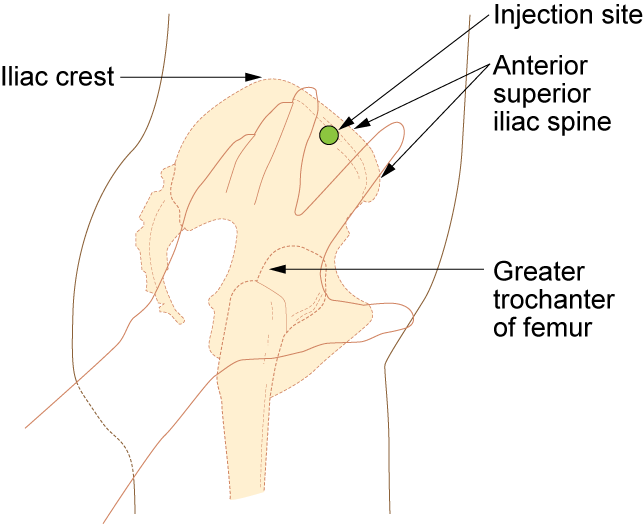
The needle gauge used at the ventrogluteal site is determined by the solution of the medication ordered. An aqueous solution can be given with a 20- to 25-gauge needle, whereas viscous or oil-based solutions are given with 18- to 21-gauge needles. The needle length is based on patient weight and body mass index. A thin adult may require a 5/8-inch to 1-inch (16 mm to 25 mm) needle, while an average adult may require a 1-inch (25 mm) needle, and a larger adult (over 70 kg) may require a 1-inch to 1½-inch (25 mm to 38 mm) needle. Children and infants require shorter needles. Refer to agency policies regarding needle length for infants, children, and adolescents. Up to 3 mL of medication may be administered in the ventrogluteal muscle of an average adult and up to 1 mL in children. See Figure 18.32[4] for an image of locating the ventrogluteal site on a patient.

Vastus Lateralis
The vastus lateralis site is commonly used for immunizations in infants and toddlers because the muscle is thick and well-developed. This muscle is located on the anterior lateral aspect of the thigh and extends from one hand’s breadth above the knee to one hand’s breadth below the greater trochanter. The outer middle third of the muscle is used for injections. To help relax the patient, ask the patient to lie flat with knees slightly bent or have the patient in a sitting position. See Figure 18.33[5] for an image of the vastus lateralis injection site.
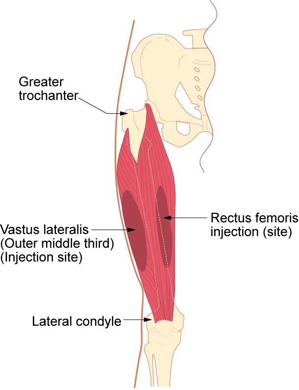
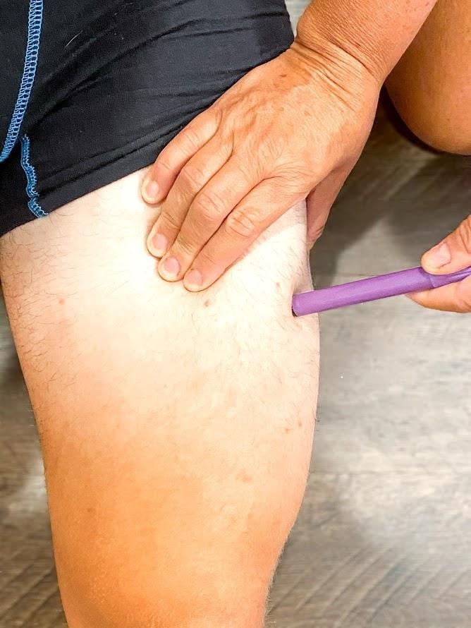
The length of the needle used at the vastus lateralis site is based on the patient’s age, weight, and body mass index. In general, the recommended needle length for an adult is 1 inch to 1 ½ inches (25 mm to 38 mm), but the needle length is shorter for children. Refer to agency policy for pediatric needle lengths. The gauge of the needle is determined by the type of medication administered. Aqueous solutions can be given with a 20- to 25-gauge needle; oily or viscous medications should be administered with 18- to 21-gauge needles. A smaller gauge needle (22 to 25 gauge) should be used with children. The maximum amount of medication for a single injection in an adult is 3 mL. See Figure 18.34[6] for an image of an intramuscular injection being administered at the vastus lateralis site.
Deltoid
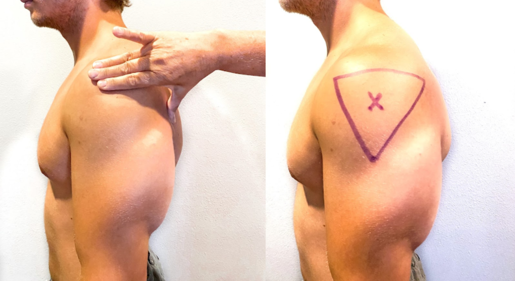
The deltoid muscle has a triangular shape and is easy to locate and access. To locate the injection site, begin by having the patient relax their arm. The patient can be standing, sitting, or lying down. To locate the landmark for the deltoid muscle, expose the upper arm and find the acromion process by palpating the bony prominence. The injection site is in the middle of the deltoid muscle, about 1 inch to 2 inches (2.5 cm to 5 cm) below the acromion process. To locate this area, lay three fingers across the deltoid muscle and below the acromion process. The injection site is generally three finger widths below in the middle of the muscle. See Figure 18.35[7] for an illustration for locating the deltoid injection site.
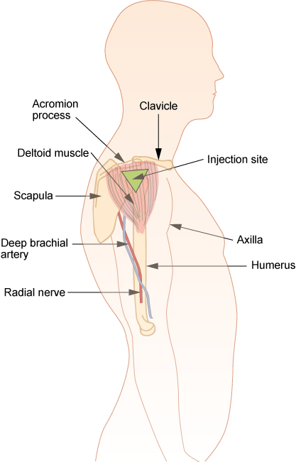
Select the needle length based on the patient’s age, weight, and body mass. In general, for an adult male weighing 60 kg to 118 kg (130 to 260 lbs), a 1-inch (25 mm) needle is sufficient. For women under 60 kg (130 lbs), a ⅝-inch (16 mm) needle is sufficient, while for women between 60 kg and 90 kg (130 to 200 lbs) a 1-inch (25 mm) needle is required. A 1 ½-inch (38 mm) length needle may be required for women over 90 kg (200 lbs) for a deltoid IM injection. For immunizations, a 22- to 25-gauge needle should be used. Refer to agency policy regarding specifications for infants, children, adolescents, and immunizations. The maximum amount of medication for a single injection is generally 1 mL. See Figure 18.36[8] for an image of locating the deltoid injection site on a patient.
Description of Procedure
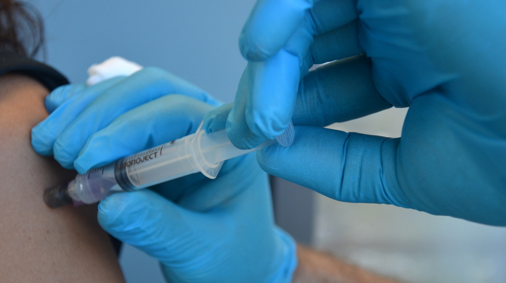
When administering an intramuscular injection, the procedure is similar to a subcutaneous injection, but instead of pinching the skin, stabilize the skin around the injection site with your nondominant hand. With your dominant hand, hold the syringe like a dart and insert the needle quickly into the muscle at a 90-degree angle using a steady and smooth motion. After the needle pierces the skin, use the thumb and forefinger of the nondominant hand to hold the syringe. If aspiration is indicated according to agency policy and manufacturer recommendations, pull the plunger back to aspirate for blood. If no blood appears, inject the medication slowly and steadily. If blood appears, discard the syringe and needle and prepare the medication again. See Figure 18.37[9] for an image of aspirating for blood. After the medication is completely injected, leave the needle in place for ten seconds, and then remove the needle using a smooth, steady motion. Remove the needle at the same angle at which it was inserted. Cover the injection site with sterile gauze using gentle pressure and apply a Band-Aid if needed.[10]
Z-track Method for IM injections
Evidence-based practice supports using the Z-track method for administration of intramuscular injections. This method prevents the medication from leaking into the subcutaneous tissue, allows the medication to stay in the muscles, and can minimize irritation.[12]
The Z-track method creates a zigzag path to prevent medication from leaking into the subcutaneous tissue. This method may be used for all injections or may be specified by the medication.
Displace the patient’s skin in a Z-track manner by pulling the skin down or to one side about 1 inch (2 cm) with your nondominant hand before administering the injection. With the skin held to one side, quickly insert the needle at a 90-degree angle. After the needle pierces the skin, continue pulling on the skin with the nondominant hand, and at the same time, grasp the lower end of the syringe barrel with the fingers of the nondominant hand to stabilize it. Move your dominant hand and pull the end of the plunger to aspirate for blood, if indicated. If no blood appears, inject the medication slowly. Once the medication is given, leave the needle in place for ten seconds. After the medication is completely injected, remove the needle using a smooth, steady motion, and then release the skin. See Figure 18.38[13] for an illustration of the Z-track method.
![“Z-track-process-1.png“ and "Z-track-process-3.png" by British Columbia Institute of Technology (BCIT) is licensed under CC BY 4.0. Access for free at https://opentextbc.ca/clinicalskills/chapter/6-8-iv-push-medications-and-saline-lock-flush/[/footnote] Illustration showing two parts of the Z track method](https://opencontent.ccbcmd.edu/app/uploads/sites/32/2024/08/ztrack-1-1024x409.png)
Special Considerations for IM Injections
- Avoid using sites with atrophied muscle because they will poorly absorb medications.
- If repeated IM injections are given, sites should be rotated to decrease the risk of hypertrophy.
- Older adults and thin patients may only tolerate up to 2 milliliters in a single injection.
- Choose a site that is free from pain, infection, abrasions, or necrosis.
- The dorsogluteal site should be avoided for intramuscular injections because of the risk for injury. If the needle inadvertently hits the sciatic nerve, the patient may experience partial or permanent paralysis of the leg.
Assessments Prior to Injection Administration
When administering any parenteral injection, the nurse assesses the patient prior to administration for safe medication administration. See Table 18.7 to review assessments prior to medication administration.
Table 18.7 Assessments Prior to Injection Administration
| Assessment | Rationale/Considerations |
|---|---|
| Check the MAR with the written medication prescription for accuracy and completeness. | Prevent medication errors. |
| Perform the rights of medication administration, including patient’s name, medication name and dose, route, time of administration, and verify the expiration date. | Prevent medication errors. Discard medication if expired. |
| Label syringes with medication names as you prepare them. | Label syringes during the procedure to administer medications safely according to The Joint Commission.[16] |
| Assess and review a patient's current medical condition, past medical history, and medication history. | Identify the need for the medication, as well as any possible contraindications for the administration of prescribed medication to the specific patient. |
| Assess the patient's history of medication allergies or nondrug allergies that may interfere with the medication. If there is a history of allergic reactions, document the type of reaction if possible (e.g., hives, rash, swelling, difficulty breathing). If there is an allergy to prescriber medication, do not prepare it and notify the prescribing provider. | Safely administer medication. |
| Review a current, evidence-based drug reference to determine medication action, indication for medication, normal dosage range, and potential side effects. Identify peak and onset times, as well as any nursing considerations. | Safely administer medication and plan to monitor the patient’s response. For example, by knowing the peak onset of fast-acting insulin, the nurse anticipates when the patient may be most at risk for a hypoglycemic reaction. |
| Review and assess pertinent laboratory results (i.e., blood glucose, partial thromboplastin time). Be aware of abnormal kidney and liver function results because they may affect metabolism of medication. Notify the prescriber of any concerns. | Collect data to determine if the medication should be withheld to ensure proper dosage and to establish a baseline for measuring the patient’s response to the drug. |
| Observe the patient’s verbal and nonverbal reactions to the injection. | Be aware of the patient’s level of anxiety and use distractions and other therapeutic techniques to reduce pain and anxiety. |
| Perform patient assessments, such as vital signs, lung sounds, or pain level, as indicated, prior to medication administration. | Obtain baseline data to ensure medication administration is appropriate at this time and to establish a baseline for measuring the patient’s response to the medication. |
| Assess for contraindications to subcutaneous or intramuscular injections such as muscle atrophy or decreased blood flow to the tissue. | Assess for contraindications because reduced muscle and blood flow interfere with the drug absorption and distribution. |
| Assess the patient’s knowledge of the medication. | Perform patient education about the medication as needed. |
| Assess the skin and tissue quality around the area of the intended injection site. Note any bruising, nonintact skin, abrasions, masses, or scar tissue. Avoid these areas and choose another recommended location. | Assess skin and tissue quality to avoid unnecessary or further injury to the already compromised skin integrity. Areas that are scarred or atrophied can affect the absorption and distribution of the medication. |
Evaluation
Nurses evaluate for possible complications of parenteral medication administration that may occur as a result of a medication error or as an adverse reaction. Complications may occur if a medication is prepared incorrectly, if the medication is injected incorrectly, or if an adverse effect occurs after the medication is injected. Unexpected outcomes can occur such as nerve or tissue damage, ineffective absorption of the medication, pain, bleeding, or infection.
An adverse reaction may develop as a result of an injected medication. An adverse reaction may also occur despite appropriate administration of medication and can happen for various reasons. A reaction may be evident within minutes or days after the exposure to the injectable medication. An unexpected outcome may range from a minor reaction, like a skin rash, to serious and life-threatening events such as anaphylaxis, hemorrhaging, and even death.
If a suspected complication occurs during administration, immediately stop the injection. Assess and monitor vital signs, notify the health care provider, and document an incident report.
Checklist for Parenteral Site Identification
Use the checklist below to review the steps for completion of "Parenteral Site Identification."
Directions: Identify parenteral injection sites, needle size/gauge, injection angle, and the appropriate amount that can be administered in each of the parenteral routes: intradermal, subcutaneous, and intramuscular.
- Describe the appropriate needle gauge, length, number of cc's, and angle for an intradermal injection:
- 25-27G
- 3/8” to 5/8”
- 0.1 mL (for TB testing)
- 5- to 15-degree angle
- Demonstrate locating the intradermal injection sites on a peer:
- Inner surface of the forearm
- Outer aspects upper arms
- Upper back below scapula
- Describe the appropriate needle gauge, length, number of mL, and angle for a subcutaneous injection:
- 25-31G
- ½” to 5/8”
- Up to 1 mL
- 45- to 90-degree angle
- Demonstrate locating the subcutaneous injection sites on a peer:
- Anterior thighs
- Abdomen
- Describe the appropriate needle gauge, length, number of mL, and angle for an adult intramuscular:
- 18-25G
- ½” - 1 ½” (based on age/size of patient and site used)
- <0.5 - 1 mL (infants and children), 2-3 mL (adults)
- 90-degree angle
- Demonstrate locating the intramuscular injection sites on a peer:
- Ventrogluteal
- Vastus lateralis
- Deltoid
- Explain how you would modify assessment techniques to reflect variations across the life span and body size variations.
Checklist for Parenteral Medication Injections
Use the checklist below to review the steps for completion of "Parenteral Medication Injections."
Steps
Disclaimer: Always review and follow agency policy regarding this specific skill.
Special Considerations:
- Plan medication administration to avoid disruption.
- Dispense medication in a quiet area.
- Avoid conversation with others.
- Follow agency's no-interruption zone policy.
- Prepare medications for ONE patient at a time.
- Plan for disposal of sharps in an appropriate sharps disposal container.
- Check the orders and MAR for accuracy and completeness; clarify any unclear orders.
- Review pertinent information related to the medications: labs, last time medication was given, and medication information: generic name, brand name, dose, route, time, class, action, purpose, side effects, contraindications, and nursing considerations.
- Gather available supplies: correctly sized syringes and needles appropriate for medication, patient's size, and site of injection; diluent (if required); tape or patient label for each syringe; nonsterile gloves; sharps container; and alcohol wipes.
- Perform hand hygiene.
- While withdrawing medication from the medication dispensing system, perform the first check of the six rights of medication administration. Check expiration date and perform any necessary calculations.
Preparing the Medication for Administration
- Scrub the top of the vial of the correct medication. State the correct dose to be drawn.
- Remove the cap from the needle. Pull back on the plunger to draw air into the syringe equal to the dose.
- With the vial on a flat surface, insert the needle. Invert the vial and withdraw the correct amount of the medication. Expel any air bubbles. Remove the needle from the vial.
- Using the scoop method, recap the needle.
- Perform the second check of the six rights of medication administration, looking at the vial, syringe, and MAR.
- Label the syringe with the name of the drug and dose.
Additional Preparation Steps When Mixing Two Types of Insulin in One Syringe: Intermediate-Acting (NPH) and Short-Acting (Regular) Insulins
- Place the vials side by side on a flat surface: NPH on left and regular insulin on the right.
- Roll the NPH insulin between your hands to mix the solution. With an alcohol pad, scrub off the vial top of the NPH insulin. Using a new alcohol pad, scrub the vial top of the regular insulin. Discard any prep pads.
- Select the correct insulin syringe that will exactly measure the TOTAL dose of the amount of NPH and regular doses (30- and 50-unit syringes measure single units; 100-unit syringes only measure even numbered doses).
- Pull back on the plunger to draw air into the syringe equal to the dose of NPH insulin.
- With the NPH vial on a flat surface, remove the cap from the syringe, insert the needle into the NPH vial, and inject air. Do not let the tip of the needle touch the insulin solution. Withdraw the needle.
- Pull back on the plunger to draw air into the syringe equal to the dose of the regular insulin.
- With the regular vial on a flat surface, insert the needle into the regular vial, and inject air.
- With the needle still in the vial, invert the regular insulin vial and withdraw the correct dose. Remove the needle from the vial. Cap the needle using the scoop method.
- Roll the NPH insulin vial between your hands to mix the solution. Uncap the needle and insert the needle into the NPH insulin vial. Withdraw the correct amount of NPH insulin. Withdraw the needle and recap using the scoop method.
- Perform the second medication check of the combined dose looking at the vial, syringe, and MAR, verifying all the rights.
- Label the syringe with the name of the combined medications and doses.
Alternative Preparation Using an Insulin Pen
- Select the correct insulin pen to be used for the injection. Identify the dose to be given.
- Remove the cap from the insulin pen and clean the top (hub) with an alcohol prep pad. Attach the insulin pen needle without contaminating the needle or pen hub.
- Turn the dial to two units and with the pen pointing upward, push the injection button to prime the pen.
- Turn the dial to the correct dose.
- Perform the second medication check looking at the insulin pen and MAR, verifying all the rights.
Administration of Parenteral Medication
- Knock, enter the room, greet the patient, and provide for privacy.
- Perform safety steps:
- Perform hand hygiene.
- Check the room for transmission-based precautions.
- Introduce yourself, your role, the purpose of your visit, and an estimate of the time it will take.
- Confirm patient ID using two patient identifiers (e.g., name and date of birth).
- Explain the process to the patient and ask if they have any questions.
- Be organized and systematic.
- Use appropriate listening and questioning skills.
- Listen and attend to patient cues.
- Ensure the patient's privacy and dignity.
- Assess ABCs.
- Perform the third check of the six rights of medication administration at the patient's bedside after performing patient identification.
- Perform the following steps according to the type of parenteral medication.
INTRADERMAL - Administration of a TB Test
- Correctly identify the sites and verbalize the landmarks used for intradermal injections.
- Select the correct site for the TB test, verbalizing the anatomical landmarks and skin considerations.
- Put on nonsterile gloves if contact with blood or body fluids is likely or if your skin or the patient's skin isn't intact.
- Use an alcohol swab in a circular motion to clean the skin at the site; place the pad above the site to mark the site, if desired.
- Gently pull the skin away from the site.
- Insert the needle with the bevel facing upward, slowly at a 5- to 15-degree angle, and then advance no more than an eighth of an inch to cover the bevel.
- Use the thumb of the nondominant hand to push on the plunger to slowly inject the medication. Inspect the site, noting if a small bleb forms under the skin surface.
- Carefully withdraw the needle straight back out of the insertion site so not to disturb the bleb (do not massage or cover the site).
- Activate the safety feature of the needle and place the syringe in the sharps container.
- Teach the patient to return for a TB skin test reading in 48-72 hours and not to press on the site or apply a Band-Aid.
SUBCUTANEOUS - Administration of Insulin in a Syringe
- Correctly identify the sites and verbalize the landmarks used for subcutaneous injections. Ask the patient regarding a preferred site of medication administration.
- Put on nonsterile gloves if contact with blood or body fluids is likely or if your skin or the patient's skin isn't intact.
- Select an appropriate site and clean with an alcohol prep in a circular motion. Place the pad above the site to mark the location, if desired. Remove the cap from the needle without contaminating the needle.
- Pinch approximately an inch of subcutaneous tissue, creating a skinfold.
- Insert the needle at a 90-degree angle, release the patient's skin, and inject the medication. Withdraw the needle.
- Activate the safety feature of the needle and place the syringe in a sharps container.
SUBCUTANEOUS - Administration with an Insulin Pen
- Select the site and clean with an alcohol prep in a circular motion. Place the pad above the site to mark the location, if desired.
- Put on nonsterile gloves if contact with blood or body fluids is likely or if your skin or the patient's skin isn't intact.
- Remove the cap from the needle without contaminating the needle.
- Pinch approximately an inch of subcutaneous tissue, creating a skinfold.
- Insert the needle quickly at a 45- to 90-degree angle (depending upon the size of the patient), continue to hold the skinfold, and inject the medication. After the medication is injected, count to 10, remove the needle, and release the skinfold.
- Dispose of the needle in a sharps container. Replace the top cap to the insulin pen.
INTRAMUSCULAR - Deltoid
- Correctly identify the site and verbalize the landmarks used for a deltoid injection.
- Put on nonsterile gloves if contact with blood or body fluids is likely or if your skin or the patient's skin isn't intact.
- Use an alcohol swab in a circular motion to clean the skin at the site. Place a pad above the site to mark the location. Remove the cap from the needle without contaminating the needle.
- Depending on the muscle mass of the deltoid, either grasp the body of the muscle between the thumb and forefingers of the nondominant hand or spread the skin taut.
- Insert the needle at a 90-degree angle.
- Follow agency policy and manufacturer recommendations regarding aspiration.
- Continue to hold the muscle fold and inject the medication. After the medication is injected, count to 10, remove the needle, and release the muscle fold.
- Activate the safety on the syringe. Place the syringe in a sharps container.
INTRAMUSCULAR - Vastus Lateralis
- Correctly identify the site and verbalize the landmarks to locate the vastus lateralis site.
- Put on nonsterile gloves if contact with blood or body fluids is likely or if your skin or the patient's skin isn't intact.
- Use an alcohol swab in a circular motion to clean the skin at the site. Place the pad above the site to mark the location. Remove the cap from the needle without contaminating the needle.
- Depending on the muscle mass of the vastus lateralis, either grasp the body of the muscle between the thumb and forefingers of the nondominant hand or spread the skin taut.
- Insert the needle at a 90-degree angle.
- Follow agency policy and manufacturer recommendations regarding aspiration.
- Continue to hold the muscle fold and inject the medication. After the medication is injected, count to 10, remove the needle, and release the muscle fold.
- Activate the safety on the syringe. Put the needle in a sharps container.
INTRAMUSCULAR - Ventrogluteal (Using the Z-track Technique)
- Correctly identify and verbalize the landmarks used to locate the ventrogluteal site.
- Put on nonsterile gloves if contact with blood or body fluids is likely or if your skin or the patient's skin isn't intact.
- Use an alcohol swab in a circular motion to clean the skin at the site and place a pad above the site to mark the location. Remove the cap from the needle without contaminating the needle.
- Place the ulnar surface of the hand approximately 1 – 3 inches from the selected site; press down and pull the skin and subcutaneous tissue to the side or downward.
- Maintaining tissue traction, hold the syringe like a dart and insert the needle into the skin at 90 degrees.
- Maintaining tissue traction, use the available thumb and index finger to help stabilize the syringe.
- Follow agency policy and manufacturer recommendations regarding aspiration. If aspiration is required, pull back the plunger and observe for blood return. If there is no blood return, inject the medication. If blood return is observed, remove the needle, and prepare a new medication.
- Maintaining tissue traction, wait 10 seconds with the needle still in the skin to allow the muscle to absorb the medication. Withdraw the needle from the site and then release traction. Do not rub/massage the site.
- Activate the safety feature of the needle; place in a sharps container.
Following Conclusion of All Injections
- Assess site; apply Band-Aid if necessary and appropriate.
- Remove gloves. Perform hand hygiene.
- Ensure safety measures when leaving the room:
- CALL LIGHT: Within reach
- BED: Low and locked (in lowest position and brakes on)
- SIDE RAILS: Secured
- TABLE: Within reach
- ROOM: Risk-free for falls (scan room and clear any obstacles)
- Document medication administered, including the site used for the injection.
View an instructor demonstration of an Intradermal Injection[17]:
View an instructor demonstration of a Subcutaneous Injection (Insulin)[18]:
View an instructor demonstration of an Intramuscular Injection[19]:
View an instructor demonstration of Using an Insulin Pen[20]:
Checklist for Parenteral Site Identification
Use the checklist below to review the steps for completion of "Parenteral Site Identification."
Directions: Identify parenteral injection sites, needle size/gauge, injection angle, and the appropriate amount that can be administered in each of the parenteral routes: intradermal, subcutaneous, and intramuscular.
- Describe the appropriate needle gauge, length, number of cc's, and angle for an intradermal injection:
- 25-27G
- 3/8” to 5/8”
- 0.1 mL (for TB testing)
- 5- to 15-degree angle
- Demonstrate locating the intradermal injection sites on a peer:
- Inner surface of the forearm
- Outer aspects upper arms
- Upper back below scapula
- Describe the appropriate needle gauge, length, number of mL, and angle for a subcutaneous injection:
- 25-31G
- ½” to 5/8”
- Up to 1 mL
- 45- to 90-degree angle
- Demonstrate locating the subcutaneous injection sites on a peer:
- Anterior thighs
- Abdomen
- Describe the appropriate needle gauge, length, number of mL, and angle for an adult intramuscular:
- 18-25G
- ½” - 1 ½” (based on age/size of patient and site used)
- <0.5 - 1 mL (infants and children), 2-3 mL (adults)
- 90-degree angle
- Demonstrate locating the intramuscular injection sites on a peer:
- Ventrogluteal
- Vastus lateralis
- Deltoid
- Explain how you would modify assessment techniques to reflect variations across the life span and body size variations.
Checklist for Parenteral Medication Injections
Use the checklist below to review the steps for completion of "Parenteral Medication Injections."
Steps
Disclaimer: Always review and follow agency policy regarding this specific skill.
Special Considerations:
- Plan medication administration to avoid disruption.
- Dispense medication in a quiet area.
- Avoid conversation with others.
- Follow agency's no-interruption zone policy.
- Prepare medications for ONE patient at a time.
- Plan for disposal of sharps in an appropriate sharps disposal container.
- Check the orders and MAR for accuracy and completeness; clarify any unclear orders.
- Review pertinent information related to the medications: labs, last time medication was given, and medication information: generic name, brand name, dose, route, time, class, action, purpose, side effects, contraindications, and nursing considerations.
- Gather available supplies: correctly sized syringes and needles appropriate for medication, patient's size, and site of injection; diluent (if required); tape or patient label for each syringe; nonsterile gloves; sharps container; and alcohol wipes.
- Perform hand hygiene.
- While withdrawing medication from the medication dispensing system, perform the first check of the six rights of medication administration. Check expiration date and perform any necessary calculations.
Preparing the Medication for Administration
- Scrub the top of the vial of the correct medication. State the correct dose to be drawn.
- Remove the cap from the needle. Pull back on the plunger to draw air into the syringe equal to the dose.
- With the vial on a flat surface, insert the needle. Invert the vial and withdraw the correct amount of the medication. Expel any air bubbles. Remove the needle from the vial.
- Using the scoop method, recap the needle.
- Perform the second check of the six rights of medication administration, looking at the vial, syringe, and MAR.
- Label the syringe with the name of the drug and dose.
Additional Preparation Steps When Mixing Two Types of Insulin in One Syringe: Intermediate-Acting (NPH) and Short-Acting (Regular) Insulins
- Place the vials side by side on a flat surface: NPH on left and regular insulin on the right.
- Roll the NPH insulin between your hands to mix the solution. With an alcohol pad, scrub off the vial top of the NPH insulin. Using a new alcohol pad, scrub the vial top of the regular insulin. Discard any prep pads.
- Select the correct insulin syringe that will exactly measure the TOTAL dose of the amount of NPH and regular doses (30- and 50-unit syringes measure single units; 100-unit syringes only measure even numbered doses).
- Pull back on the plunger to draw air into the syringe equal to the dose of NPH insulin.
- With the NPH vial on a flat surface, remove the cap from the syringe, insert the needle into the NPH vial, and inject air. Do not let the tip of the needle touch the insulin solution. Withdraw the needle.
- Pull back on the plunger to draw air into the syringe equal to the dose of the regular insulin.
- With the regular vial on a flat surface, insert the needle into the regular vial, and inject air.
- With the needle still in the vial, invert the regular insulin vial and withdraw the correct dose. Remove the needle from the vial. Cap the needle using the scoop method.
- Roll the NPH insulin vial between your hands to mix the solution. Uncap the needle and insert the needle into the NPH insulin vial. Withdraw the correct amount of NPH insulin. Withdraw the needle and recap using the scoop method.
- Perform the second medication check of the combined dose looking at the vial, syringe, and MAR, verifying all the rights.
- Label the syringe with the name of the combined medications and doses.
Alternative Preparation Using an Insulin Pen
- Select the correct insulin pen to be used for the injection. Identify the dose to be given.
- Remove the cap from the insulin pen and clean the top (hub) with an alcohol prep pad. Attach the insulin pen needle without contaminating the needle or pen hub.
- Turn the dial to two units and with the pen pointing upward, push the injection button to prime the pen.
- Turn the dial to the correct dose.
- Perform the second medication check looking at the insulin pen and MAR, verifying all the rights.
Administration of Parenteral Medication
- Knock, enter the room, greet the patient, and provide for privacy.
- Perform safety steps:
- Perform hand hygiene.
- Check the room for transmission-based precautions.
- Introduce yourself, your role, the purpose of your visit, and an estimate of the time it will take.
- Confirm patient ID using two patient identifiers (e.g., name and date of birth).
- Explain the process to the patient and ask if they have any questions.
- Be organized and systematic.
- Use appropriate listening and questioning skills.
- Listen and attend to patient cues.
- Ensure the patient's privacy and dignity.
- Assess ABCs.
- Perform the third check of the six rights of medication administration at the patient's bedside after performing patient identification.
- Perform the following steps according to the type of parenteral medication.
INTRADERMAL - Administration of a TB Test
- Correctly identify the sites and verbalize the landmarks used for intradermal injections.
- Select the correct site for the TB test, verbalizing the anatomical landmarks and skin considerations.
- Put on nonsterile gloves if contact with blood or body fluids is likely or if your skin or the patient's skin isn't intact.
- Use an alcohol swab in a circular motion to clean the skin at the site; place the pad above the site to mark the site, if desired.
- Gently pull the skin away from the site.
- Insert the needle with the bevel facing upward, slowly at a 5- to 15-degree angle, and then advance no more than an eighth of an inch to cover the bevel.
- Use the thumb of the nondominant hand to push on the plunger to slowly inject the medication. Inspect the site, noting if a small bleb forms under the skin surface.
- Carefully withdraw the needle straight back out of the insertion site so not to disturb the bleb (do not massage or cover the site).
- Activate the safety feature of the needle and place the syringe in the sharps container.
- Teach the patient to return for a TB skin test reading in 48-72 hours and not to press on the site or apply a Band-Aid.
SUBCUTANEOUS - Administration of Insulin in a Syringe
- Correctly identify the sites and verbalize the landmarks used for subcutaneous injections. Ask the patient regarding a preferred site of medication administration.
- Put on nonsterile gloves if contact with blood or body fluids is likely or if your skin or the patient's skin isn't intact.
- Select an appropriate site and clean with an alcohol prep in a circular motion. Place the pad above the site to mark the location, if desired. Remove the cap from the needle without contaminating the needle.
- Pinch approximately an inch of subcutaneous tissue, creating a skinfold.
- Insert the needle at a 90-degree angle, release the patient's skin, and inject the medication. Withdraw the needle.
- Activate the safety feature of the needle and place the syringe in a sharps container.
SUBCUTANEOUS - Administration with an Insulin Pen
- Select the site and clean with an alcohol prep in a circular motion. Place the pad above the site to mark the location, if desired.
- Put on nonsterile gloves if contact with blood or body fluids is likely or if your skin or the patient's skin isn't intact.
- Remove the cap from the needle without contaminating the needle.
- Pinch approximately an inch of subcutaneous tissue, creating a skinfold.
- Insert the needle quickly at a 45- to 90-degree angle (depending upon the size of the patient), continue to hold the skinfold, and inject the medication. After the medication is injected, count to 10, remove the needle, and release the skinfold.
- Dispose of the needle in a sharps container. Replace the top cap to the insulin pen.
INTRAMUSCULAR - Deltoid
- Correctly identify the site and verbalize the landmarks used for a deltoid injection.
- Put on nonsterile gloves if contact with blood or body fluids is likely or if your skin or the patient's skin isn't intact.
- Use an alcohol swab in a circular motion to clean the skin at the site. Place a pad above the site to mark the location. Remove the cap from the needle without contaminating the needle.
- Depending on the muscle mass of the deltoid, either grasp the body of the muscle between the thumb and forefingers of the nondominant hand or spread the skin taut.
- Insert the needle at a 90-degree angle.
- Follow agency policy and manufacturer recommendations regarding aspiration.
- Continue to hold the muscle fold and inject the medication. After the medication is injected, count to 10, remove the needle, and release the muscle fold.
- Activate the safety on the syringe. Place the syringe in a sharps container.
INTRAMUSCULAR - Vastus Lateralis
- Correctly identify the site and verbalize the landmarks to locate the vastus lateralis site.
- Put on nonsterile gloves if contact with blood or body fluids is likely or if your skin or the patient's skin isn't intact.
- Use an alcohol swab in a circular motion to clean the skin at the site. Place the pad above the site to mark the location. Remove the cap from the needle without contaminating the needle.
- Depending on the muscle mass of the vastus lateralis, either grasp the body of the muscle between the thumb and forefingers of the nondominant hand or spread the skin taut.
- Insert the needle at a 90-degree angle.
- Follow agency policy and manufacturer recommendations regarding aspiration.
- Continue to hold the muscle fold and inject the medication. After the medication is injected, count to 10, remove the needle, and release the muscle fold.
- Activate the safety on the syringe. Put the needle in a sharps container.
INTRAMUSCULAR - Ventrogluteal (Using the Z-track Technique)
- Correctly identify and verbalize the landmarks used to locate the ventrogluteal site.
- Put on nonsterile gloves if contact with blood or body fluids is likely or if your skin or the patient's skin isn't intact.
- Use an alcohol swab in a circular motion to clean the skin at the site and place a pad above the site to mark the location. Remove the cap from the needle without contaminating the needle.
- Place the ulnar surface of the hand approximately 1 – 3 inches from the selected site; press down and pull the skin and subcutaneous tissue to the side or downward.
- Maintaining tissue traction, hold the syringe like a dart and insert the needle into the skin at 90 degrees.
- Maintaining tissue traction, use the available thumb and index finger to help stabilize the syringe.
- Follow agency policy and manufacturer recommendations regarding aspiration. If aspiration is required, pull back the plunger and observe for blood return. If there is no blood return, inject the medication. If blood return is observed, remove the needle, and prepare a new medication.
- Maintaining tissue traction, wait 10 seconds with the needle still in the skin to allow the muscle to absorb the medication. Withdraw the needle from the site and then release traction. Do not rub/massage the site.
- Activate the safety feature of the needle; place in a sharps container.
Following Conclusion of All Injections
- Assess site; apply Band-Aid if necessary and appropriate.
- Remove gloves. Perform hand hygiene.
- Ensure safety measures when leaving the room:
- CALL LIGHT: Within reach
- BED: Low and locked (in lowest position and brakes on)
- SIDE RAILS: Secured
- TABLE: Within reach
- ROOM: Risk-free for falls (scan room and clear any obstacles)
- Document medication administered, including the site used for the injection.
View an instructor demonstration of an Intradermal Injection[21]:
View an instructor demonstration of a Subcutaneous Injection (Insulin)[22]:
View an instructor demonstration of an Intramuscular Injection[23]:
View an instructor demonstration of Using an Insulin Pen[24]:
Patient reports post-surgical pain at a level of 8/10. Patient is grimacing when moving in the bed and describes pain as a dull constant ache located in the right lower abdomen surgical incision area that is aggravated by moving or repositioning. Blood pressure 146/80, heart rate 94 bpm, respirations 22 per minute, temperature (tympanic) 98.8°F, pulse oximetry 98%. Last pain medication Ketorolac 30 mg (IM) given 8 hours ago. Incision site is dry and intact. Ketorolac 30mg IM administered per health provider’s prescription in the left ventrogluteal area with 22-gauge needle, length 1 ½ inches. Patient tolerated the procedure without difficulty or increased pain. Injection site post-procedure without bleeding or hematoma.
Patient reports post-surgical pain at a level of 8/10. Patient is grimacing when moving in the bed and describes pain as a dull constant ache located in the right lower abdomen surgical incision area that is aggravated by moving or repositioning. Blood pressure 146/80, heart rate 94 bpm, respirations 22 per minute, temperature (tympanic) 98.8°F, pulse oximetry 98%. Last pain medication Ketorolac 30 mg (IM) given 8 hours ago. Incision site is dry and intact. Ketorolac 30mg IM administered per health provider’s prescription in the left ventrogluteal area with 22-gauge needle, length 1 ½ inches. Patient tolerated the procedure without difficulty or increased pain. Injection site post-procedure without bleeding or hematoma.

