7.2 Review of Anatomy and Physiology of the Endocrine System
Open Resources for Nursing (Open RN)
The endocrine system includes several glands, including the pineal, hypothalamus, pituitary, thyroid, parathyroid, and adrenal glands, as well as the pancreas, ovaries, and testes. See Figure 7.1 for an illustration of these organs of the endocrine system.[1]
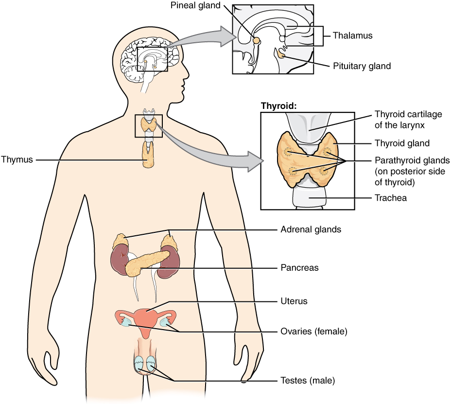
Endocrine glands secrete hormones as chemical signaling. Hormones are transported via the bloodstream throughout the body, where they bind to receptors on target cells, triggering a characteristic response. This long-distance communication is the fundamental function of the endocrine system.
Pineal Gland
The pineal gland is a small cone-shaped structure that extends posteriorly from a ventricle of the brain. The pineal gland produces the hormone melatonin and secretes it directly into the cerebrospinal fluid, which carries it into the blood. Melatonin affects reproductive development and daily circadian rhythms.[2]
Thymus Gland
The thymus gland is located in the mediastinum. It is responsible for production of T lymphocytes for the body’s immune response.
Hypothalamus and Pituitary
The hypothalamus can be viewed as the body’s control center. The hypothalamus connects to the pituitary gland by the stalk-like infundibulum. The pituitary gland is about the size of a pea and consists of an anterior and posterior lobe. Each secretes different hormones in response to signals from the hypothalamus. See Figure 7.2 for an illustration of the hypothalamus–pituitary complex.[3]
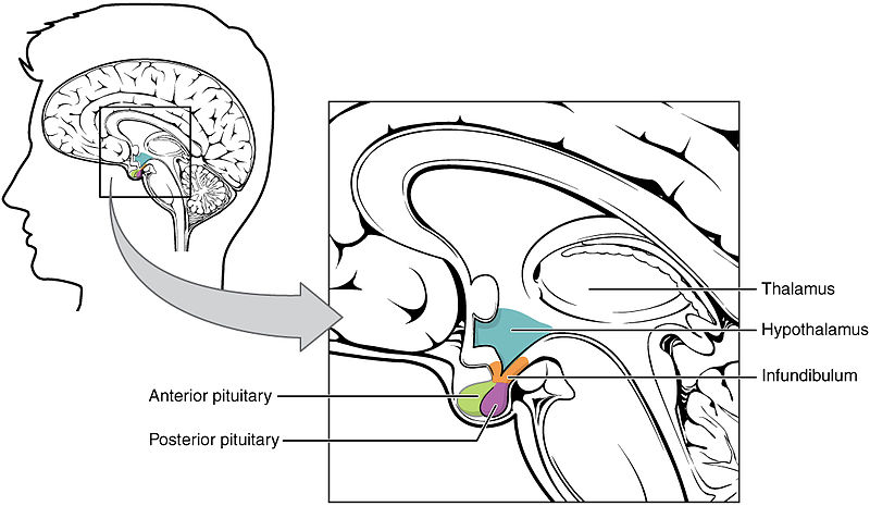
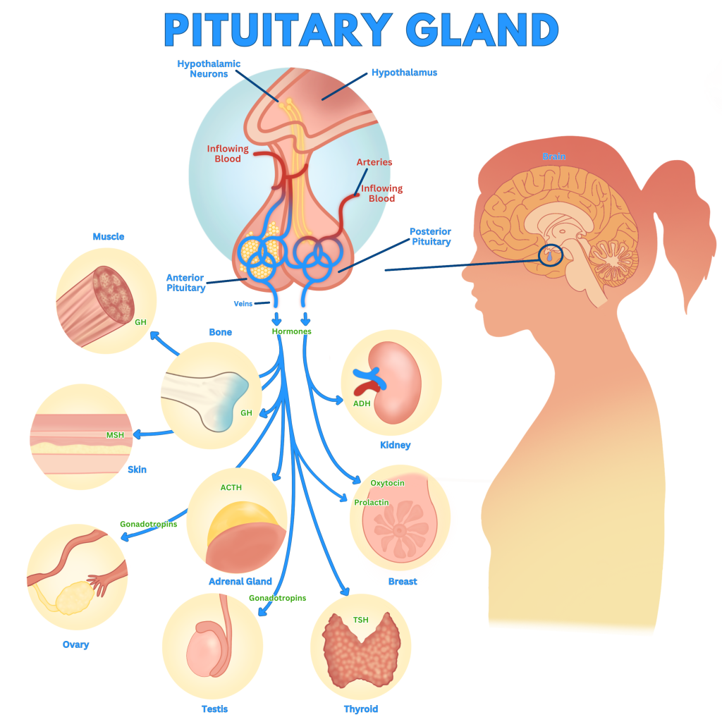
The hypothalamus–pituitary complex releases hormones that stimulate secretion of other hormones by other endocrine glands and also produce direct responses in target tissues. Its main function is to maintain a stable state called homeostasis. See Figure 7.3[4] for an illustration of the effects of the hormones released by the anterior and posterior pituitary glands.
Posterior Pituitary
The posterior pituitary gland secretes two hormones produced by the hypothalamus called oxytocin and antidiuretic hormone (ADH).
- Oxytocin stimulates labor contractions and lactation after delivery.
- Antidiuretic hormone (ADH) regulates blood osmolarity by targeting the kidneys to increase water reabsorption.
Blood osmolarity refers to the concentration of sodium and other solutes in the blood. Blood osmolarity levels change in response to fluid intake, salty food ingestion, head injuries, disease, and side effects of medications. Blood osmolarity is constantly monitored by osmoreceptors in the hypothalamus.
As blood osmolarity increases beyond normal levels, the posterior pituitary is stimulated to release ADH. For example, high blood osmolarity can occur if someone eats a very salty meal or becomes dehydrated from inadequate intake of fluids. Osmoreceptors sense the high blood osmolarity and trigger the sensation of thirst, while also stimulating the posterior pituitary to release ADH. ADH targets the kidneys to increase water reabsorption. As more water is reabsorbed by the kidneys and is returned to the blood, blood osmolarity decreases.
In contrast, if blood osmolarity decreases beyond normal levels, the release of ADH is inhibited, causing the kidney to increase the elimination of water and resulting in dilute urine.
Drugs can also affect the secretion of ADH or imitate its effects. For example, alcohol consumption inhibits the release of ADH, resulting in increased dilute urine production that can eventually lead to dehydration (and a hangover). Another example is vasopressin, a synthetic ADH medication that is used to treat very low blood pressure by causing the kidneys to retain water. Vasopressin is also used to treat a disease called diabetes insipidus (DI). People with DI have decreased amounts of ADH released by their pituitary, resulting in excessive amounts of dilute urine and severe dehydration.[5]
Anterior Pituitary
The anterior pituitary produces seven hormones:
- Growth hormone (GH): Promotes growth in children, helps maintain normal body structure in adults, and plays a role in metabolism in both children and adults.
- Thyroid-stimulating hormone (TSH): Stimulates thyroid hormone release by the thyroid gland. For example, if thyroid hormone levels in the blood are too low, the pituitary gland makes larger amounts of TSH to tell the thyroid to work harder. Conversely, if thyroid hormone levels are too high, the pituitary gland releases little or no TSH.
- Adrenocorticotropic hormone (ACTH): Regulates the release of glucocorticoid hormone (cortisol) by the adrenal gland.
- Follicle-stimulating hormone (FSH): Plays a role in sexual development and reproduction by affecting the function of the ovaries and testes.
- Luteinizing hormone (LH): Plays a role in sexual development in children and in women and triggers ovulation (the release of an egg from the ovary) during the menstrual cycle.
- Beta-endorphin: Possesses morphine-like effects and plays a role in pain management and natural reward circuits such as feeding, drinking, sex, and maternal behavior.
- Prolactin: Stimulates breast development and milk production in females.
Of the hormones released by the anterior pituitary, TSH, ACTH, FSH, and LH are referred to as tropic hormones because they turn on or off the function of other endocrine glands.
Negative Feedback Loop
TSH and ACTH levels in the blood are controlled by a negative feedback loop with the hypothalamus.
Thyroid-Stimulating Hormone (TSH)
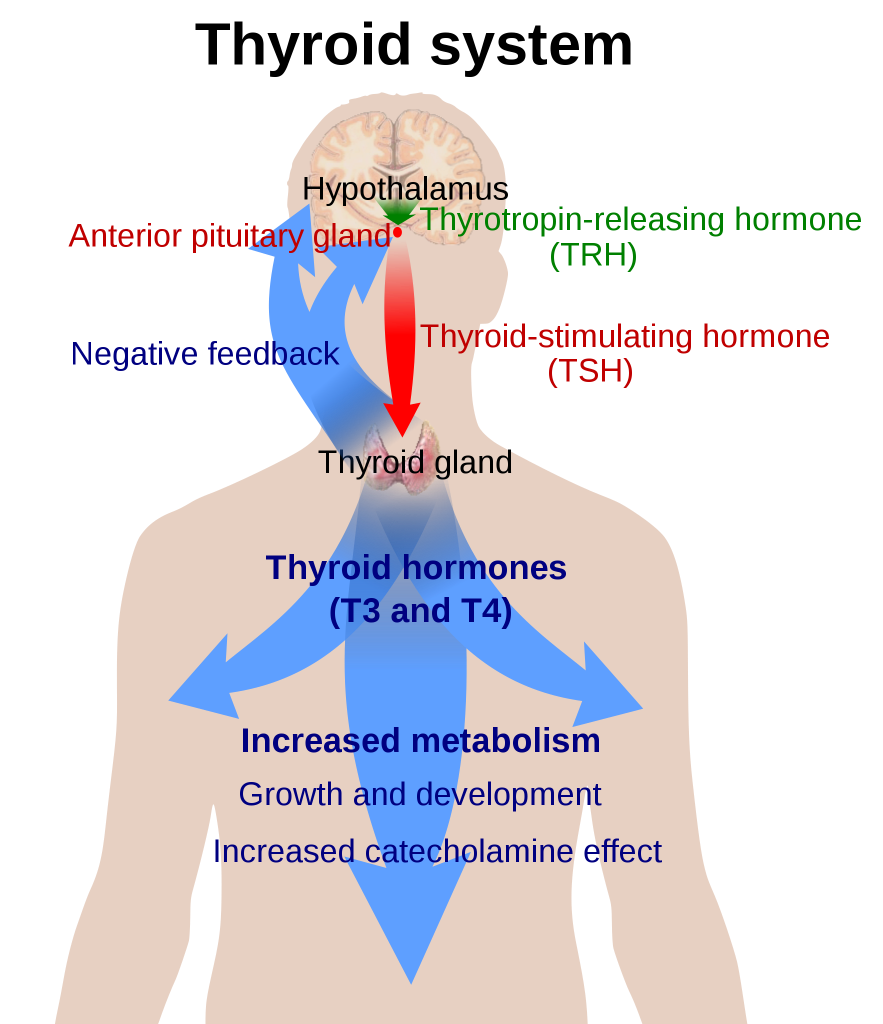
The hypothalamus stimulates the anterior pituitary with thyrotropin-releasing hormone (TRH). In response, the anterior pituitary stimulates the thyroid with thyroid-stimulating hormone (TSH). In response to TSH, the thyroid releases thyroid hormones called T3 and T4.
A negative feedback loop regulates the release of TSH by the anterior pituitary. For example, if the level of thyroid hormones (T3 and T4) decreases in the bloodstream, the hypothalamus increases the secretion of TRH. TRH causes the anterior pituitary to increase the secretion of TSH. TSH stimulates the thyroid to increase production and release of T3 and T4. For this reason, a hypofunctioning thyroid is often diagnosed by elevated TSH levels. See Figure 7.4[6] for an image of the hypothalamus-anterior pituitary-thyroid axis and the corresponding negative feedback loop.
Adrenocorticotropic Hormone (ACTH)
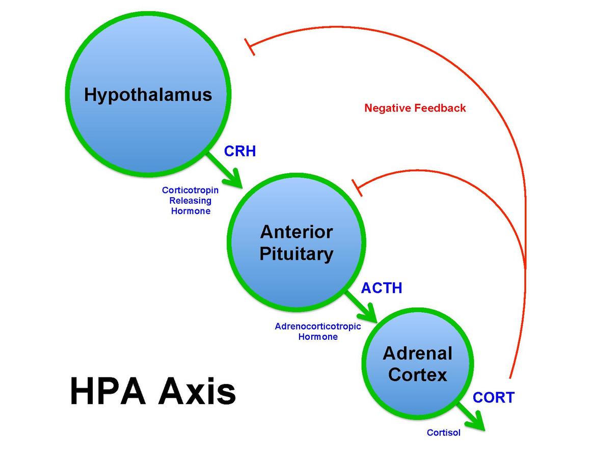
The hypothalamus stimulates the anterior pituitary with corticotropin-releasing hormone (CRH). In response, the anterior pituitary releases adrenocorticotropic hormone (ACTH) to stimulate the adrenal cortex to release corticosteroids.[7]
A negative feedback loop regulates the release of ACTH by the anterior pituitary. For example, if levels of corticosteroids decrease in the bloodstream, the hypothalamus increases the secretion of CRH, causing the anterior pituitary to increase the secretion of ACTH. This is often referred to as the hypothalamus-pituitary-adrenal (HPA) axis. See Figure 7.5[8] for an image of the HPA axis and negative feedback loop.
Thyroid Gland
The thyroid gland is a butterfly-shaped endocrine gland located in the neck, just below the larynx. It helps to regulate metabolic processes in the body by producing and releasing thyroid hormones.
The thyroid releases these two hormones:
- Thyroxine (T4)
- Triiodothyronine (T3)
Both T3 and T4 are iodine-containing compounds. These hormones are critical for regulating the body’s basal metabolic rate (BMR), the rate at which the body burns energy at rest.
The primary functions of thyroid hormones T3 and T4 include the following:
- Regulation of Metabolism: Control how the body uses energy and oxygen, impacting processes such as digestion, heart rate, and temperature regulation.
- Growth and Development: Impact normal growth and development, particularly in children and infants. They influence the growth of bones, as well as the development of the brain and the nervous system.
- Temperature Regulation: Regulate body temperature by controlling heat production and heat dissipation mechanisms.
- Energy Production: Involved in the conversion of food into energy. They affect the breakdown of carbohydrates, fats, and proteins for energy use.
- Heart Rate and Blood Pressure: Influence heart rate and the strength of heart contractions, helping to maintain cardiovascular function.
- Brain Function: Play a role in cognitive function, mood regulation, and mental alertness. For example, hypothyroidism (low thyroid hormone levels) can lead to cognitive and emotional changes.
The production and release of thyroid hormones are regulated by the hypothalamus-pituitary complex discussed earlier in this chapter.
Parathyroid Glands
Four small masses of tissue are embedded on the surface of the thyroid gland called parathyroid glands.
Parathyroid glands secrete parathyroid hormone (PTH). PTH maintains homeostasis between calcium and phosphorus via a negative feedback loop. Calcium and phosphorus have an inverse relationship, meaning if phosphorus levels rise, calcium levels will drop.
Adrenal Glands
The adrenal glands are small glands located on top of each kidney. There are two parts to the adrenal gland called the adrenal cortex and the adrenal medulla. Each part has distinct functions:
- Adrenal Cortex: The adrenal cortex is responsible for producing a group of steroid hormones known as corticosteroids. Corticosteroids can be further divided into three categories called mineralocorticoids, glucocorticoids, and androgens.
- Mineralocorticoids: The principal mineralocorticoid is aldosterone. Aldosterone regulates electrolyte and fluid balance in the body by controlling sodium and potassium levels in the blood. It is a key component of the renin-angiotensin-aldosterone system (RAAS) in which specialized cells of the kidneys secrete renin in response to low blood volume or low blood pressure. Renin then catalyzes angiotensinogen to the hormone Angiotensin I. Angiotensin I is converted to Angiotensin II by the angiotensin-converting enzyme (ACE). Angiotensin II stimulates the release of aldosterone. Many cardiac medications target the effects of aldosterone and the RAAS system. For example, ACE inhibitors block the production of Angiotensin II and are used to reduce high blood pressure.
- Glucocorticoids: The principal glucocorticoid is cortisol. Cortisol is the most important glucocorticoid because it plays a crucial role in metabolism, immune response, and the body’s response to stress. Cortisol helps regulate blood sugar levels, suppress inflammation, and manage the stress response.
- Androgens: These are secreted in minimal amounts in both sexes by the adrenal cortex, but their effect is usually masked by the hormones from the testes and ovaries.
- Adrenal Medulla: The adrenal medulla is responsible for producing epinephrine and norepinephrine. These hormones play a central role in the body’s “fight or flight” response to stress that is triggered by the sympathetic nervous system. Epinephrine and norepinephrine prepare the body for action by increasing heart rate, dilating airways, and redirecting blood flow to muscles, among other effects.
View a supplementary YouTube video[9] on ACTH and the adrenal gland:
Ovaries and Testes
In females, the ovaries produce ova (eggs), and in males, the testes produce sperm. Ovaries and testes also secrete hormones.
Ovaries secrete estrogen and progesterone[10]:
- At the onset of puberty, estrogen promotes the development of breasts and the maturation of the uterus.
- Progesterone causes the uterine lining to thicken in preparation for pregnancy.
- Together, progesterone and estrogen are responsible for the changes that occur in the uterus during the female menstrual cycle.
Testes secrete testosterone. At the onset of puberty, testosterone is responsible for the following actions[11]:
- The growth and development of the male reproductive structures
- Increased skeletal and muscular growth
- Enlargement of the larynx accompanied by voice changes
- Growth and distribution of body hair
Pancreas
The pancreas is a long, flat gland that lies behind the stomach. The pancreas serves two roles called exocrine and endocrine. The exocrine role refers to the release of digestive enzymes called amylase and lipase that help to digest food. The endocrine role refers to the production of glucagon and insulin by clusters of cells called islet cells to regulate blood glucose levels and keep them within a healthy range. See Figure 7.6[12] for an illustration of the pancreas.
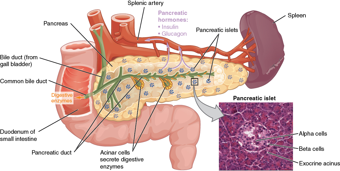
The islet cells in the pancreas include alpha, beta, and delta cells:
- Alpha Cells: Alpha cells secrete a hormone called glucagon. The primary function of glucagon is to increase blood glucose levels. When blood glucose levels drop (such as between meals or during exercise), glucagon is released.
- Beta Cells: Beta cells secrete insulin. Insulin facilitates the uptake of glucose by cells from the bloodstream, thus reducing blood glucose levels.
- Delta Cells: Delta cells secrete somatostatin. Somatostatin slows down the secretion of insulin and glucagon when needed to maintain blood glucose homeostasis.
Regulation of Blood Glucose Levels by Insulin and Glucagon
Glucose is the primary fuel for all body cells and the only fuel source for the brain. The digestive system breaks down carbohydrates into glucose, where it is absorbed into the bloodstream. Glucose is taken up by cells for fuel. Glucose that is not immediately used by cells is stored in the liver and muscles as glycogen. It is also converted into triglycerides and stored in adipose (fat) tissue.
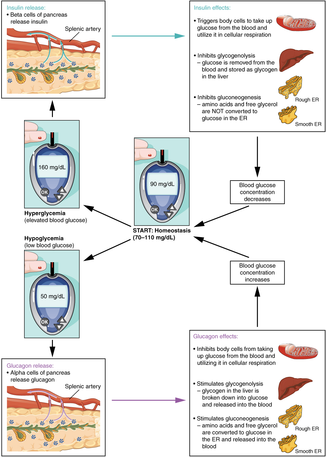
Blood glucose levels are maintained by healthy individuals’ bodies between 70 mg/dL and 110 mg/dL. Receptors located in the pancreas sense blood glucose levels, and the islet cells secrete glucagon or insulin to maintain normal blood glucose levels. If blood glucose levels rise above this range, insulin is released, which stimulates body cells to take in glucose from the blood. If blood glucose levels drop below this range, glucagon is released, which stimulates cells to release glucose from the stored glycogen into the bloodstream.
Glucagon
When blood glucose levels drop between meals or during exercise, the alpha cells secrete glucagon, which increases blood glucose levels by stimulating the following actions:
- Within cells, it inhibits the uptake of glucose.
- It stimulates the liver to convert stored glycogen into glucose and release it into the bloodstream, a process called glycogenolysis.
- It stimulates the liver to take up amino acids from the blood and converts them into glucose, a process called gluconeogenesis.
- It stimulates the breakdown of stored triglycerides into free fatty acids and glycerol, a process called lipolysis. Glycerol travels to the liver, where it is converted into glucose. This is also a form of gluconeogenesis.
Insulin
The presence of food in the intestine triggers the beta cells in the pancreas to produce and secrete insulin. As blood glucose levels continue to rise as nutrients are absorbed into the bloodstream, insulin continues to be released and stimulates the following actions:
- It stimulates the uptake of glucose by cells by triggering the movement of glucose transporters to the cell membranes, which move glucose into the interior of cells for energy. Glycolysis is the first step in the breakdown of glucose to extract energy for cellular metabolism.
- It stimulates the liver to convert excess blood glucose into glycogen for storage. It also inhibits glycogenolysis and gluconeogenesis.
- Excess glucose is synthesized into triglycerides, a process called lipogenesis.
The secretion of insulin and glucagon by the pancreas is regulated through a negative feedback loop. As glucose levels decrease in the bloodstream, insulin release is inhibited. Conversely, as glucose levels rise in the bloodstream, glucagon release is inhibited. See Figure 7.7[13] for an illustration of the homeostatic regulation of blood glucose levels.
- “1801 The Endocrine System.jpg” by OpenStax is licensed under CC BY 3.0 ↵
- National Cancer Institute. (n.d.). Endocrine glands & their function. https://training.seer.cancer.gov/anatomy/endocrine/glands/ ↵
- “1806 The Hypothalamus-Pituitary Complex.jpg” by OpenStax is licensed under CC BY 3.0 ↵
- "Pituitary Gland" by Meredith Pomietlo is licensed under CC BY 4.0 ↵
- This work is a derivative of Anatomy and Physiology by OpenStax licensed under CC BY 4.0. Access for free at https://openstax.org/books/anatomy-and-physiology/pages/1-introduction ↵
- “Thyroid_system.svg” by Mikael Häggström is licensed in the Public Domain. ↵
- This work is a derivative of Anatomy and Physiology by OpenStax licensed under CC BY 4.0. Access for free at https://openstax.org/books/anatomy-and-physiology/pages/1-introduction ↵
- “Brian_M_Sweis_HPA_Axis_Diagram_2012.pdf” by BrianMSweis is licensed under CC BY-SA 3.0 ↵
- Forciea, B. (2015, May 12). Anatomy and physiology: Endocrine system: ACTH (Adrenocorticotropin hormone) V2.0 [Video]. YouTube. All rights reserved. Video reused with permission. https://youtu.be/4m7XflJzm2w. ↵
- National Cancer Institute. (n.d.). Gonads. https://training.seer.cancer.gov/anatomy/endocrine/glands/gonads.html ↵
- National Cancer Institute. (n.d.). Gonads. https://training.seer.cancer.gov/anatomy/endocrine/glands/gonads.html ↵
- “1820 The Pancreas.jpg” by OpenStax is licensed under CC BY 3.0 ↵
- “1822 The Homeostatic Regulation of Blood Glucose Levels.jpg” by OpenStax is licensed under CC BY 3.0 ↵
A lifelong problem-solving approach that integrates the best evidence from well-designed research studies and evidence-based theories; clinical expertise and evidence from assessment of the health care consumer’s history and condition, as well as health care resources; and patient, family, group, community, and population preferences and values.
Conditions in the places where people live, learn, work, and play, such as unstable housing, low income areas, unsafe neighborhoods, or substandard education that affect a wide range of health risks and outcomes.
Personal values, character, or conduct of individuals or groups within communities and societies.
The belief that one’s culture (or race, ethnicity, or country) is better and preferable than another’s.
Personal values, character, or conduct of individuals or groups within communities and societies.
The prevailing standards of behavior of a society that enable people to live cooperatively in groups.
An ethical theory based on rules that distinguish right from wrong.
An ethical theory based on rules that distinguish right from wrong.
Principles used to define nurses’ moral duties and aid in ethical analysis and decision-making.
The capacity to determine one’s own actions through independent choice, including demonstration of competence.
The act or process of pleading for, supporting, or recommending a cause or course of action.
Feelings occurring when correct ethical action is identified but the individual feels constrained by competing values of an organization or other individuals.
A formal committee established by a health care organization to problem-solve ethical dilemmas.
A formal committee established by a health care organization to problem-solve ethical dilemmas.
The exam that nursing graduates must pass successfully to obtain their nursing license and become a registered nurse.
A concise summary of the content and scope of the NCLEX that serves as an excellent guide for preparing for the exam. NCLEX-RN test plans are updated every three years based on surveys of newly licensed registered nurses to ensure the NCLEX questions reflect fair, comprehensive, current, and entry-level nursing competency.
The process by which a State Board of Nursing (SBON) grants permission to an individual to engage in nursing practice after verifying the applicant has attained the competency necessary to perform the scope of practice of a registered nurse (RN).
State legislation that allows nurses to practice in other NLC states with their original state’s nursing license without having to obtain additional licenses, contingent upon remaining a resident of that state.
A permit issued by the State Board of Nursing (SBON) that allows the applicant to practice practical nursing under the direct supervision of a registered nurse until their RN license is granted.
A document that highlights one’s background, education, skills, and accomplishments to potential employers.
A document that highlights one’s background, education, skills, and accomplishments to potential employers.
A compilation of materials showcasing examples of previous work demonstrating one’s skills, qualifications, education, training, and experience.
Learning Activities
(Answers to "Learning Activities" can be found in the '"Answer Key'" at the end of the book. Answers to interactive activity elements will be provided within the element as immediate feedback.)
1. A male patient has an impairment of cranial nerve II. Specific to this impairment, the nurse would plan to do which of the following to ensure patient safety?
- Use a loud tone when speaking to the patient
- Test the temperature of the shower water
- Check the temperature of the food prior to eating
- Remove obstacles when ambulating
2. The nurse is performing a mental status examination on a patient with confusion. This test assesses which of the following?
- Cerebral function
- Cerebellar function
- Sensory function
- Intellectual function
Test your clinical judgment with an NCLEX Next Generation-style question: Chapter 6, Assignment 1.
Test your clinical judgment with an NCLEX Next Generation-style question: Chapter 6, Assignment 2.
Experienced and competent RNs who serve as a role model and a resource to a newly hired nurse.
Experienced and competent RNs who serve as a role model and a resource to a newly hired nurse.
A transition process that provides additional professional development for newly licensed nurses.
A transition process that provides additional professional development for newly licensed nurses.
A theory by Dr. Patricia Benner that explains how new hires develop skills and a holistic understanding of patient care over time, resulting from a combination of a strong educational foundation and thorough clinical experiences.
A theory by Dr. Patricia Benner that explains how new hires develop skills and a holistic understanding of patient care over time, resulting from a combination of a strong educational foundation and thorough clinical experiences.
National standards of care and treatment processes for common conditions. These processes are proven to reduce complications and lead to better patient outcomes.
The degree to which nursing services for health care consumers, families, groups, communities, and populations increase the likelihood of desirable outcomes and are consistent with evolving nursing knowledge.
The degree to which nursing services for health care consumers, families, groups, communities, and populations increase the likelihood of desirable outcomes and are consistent with evolving nursing knowledge.
A review process to determine if an agency meets the defined standards of quality determined by the accrediting body.
A review process to determine if an agency meets the defined standards of quality determined by the accrediting body.
National standards of care and treatment processes for common conditions. These processes are proven to reduce complications and lead to better patient outcomes.
Guidelines specific to organizations accredited by The Joint Commission that focus on problems in health care safety and ways to solve them.
An investigation by insurance agencies and other health care funders on services performed by doctors, nurses, and other health care team members to ensure money is not wasted covering things that are unnecessary for proper treatment or are inefficient.
An investigation by insurance agencies and other health care funders on services performed by doctors, nurses, and other health care team members to ensure money is not wasted covering things that are unnecessary for proper treatment or are inefficient.
Using information and technology to communicate, manage knowledge, mitigate error, and support decision-making.
Using information and technology to communicate, manage knowledge, mitigate error, and support decision-making.
A systematic process using measurable data to improve health care services and the overall health status of patients.
The science and practice integrating nursing, its information and knowledge, with information and communication technologies to promote the health of people, families, and communities worldwide.
The science and practice integrating nursing, its information and knowledge, with information and communication technologies to promote the health of people, families, and communities worldwide.
A systematic process using measurable data to improve health care services and the overall health status of patients.
Provide objective data by using number values to explain outcomes.
Provide objective data by using number values to explain outcomes.
Provide subjective data, often focusing on the perception or experience of the participants.
Provide subjective data, often focusing on the perception or experience of the participants.
Reviews other independent research studies asking similar research questions.
