5.3 General Cardiovascular System Assessment
Open Resources for Nursing (Open RN)
When evaluating a client for possible disorders of the cardiovascular system, the nurse must attend to individual and population risk factors, cultural influence, and socioeconomic factors that may impact preventative and curative health. Client health history, physical examination findings, and diagnostic test results will also play an important role in cardiovascular system assessment.
Risk Factors, Cultural, and Socioeconomic Influences on Cardiovascular System Health
Risk Factors
Cardiovascular disease (CVD) remains a significant global health concern, requiring providers to conduct a comprehensive risk assessment to guide client prevention and management strategies. A thorough client history is foundational for evaluating CVD risk, encompassing both modifiable and nonmodifiable risk factors. Understanding contributing risk factors provides essential insights into understanding an individual’s susceptibility to developing CVD and helps health care professionals tailor interventions effectively.
In the context of client history, identifying modifiable risk factors is crucial. Modifiable risk factors are risk factors that an individual can control through their actions, behavior, or lifestyle choices. These include factors such as tobacco use, physical activity, dietary habits, stress management, and sleep habits. Metabolic syndrome is a group of conditions that increases a person’s risk of coronary heart disease, diabetes, and stroke. The conditions include elevated blood pressure, blood glucose, and triglyceride levels, as well as increased waist circumference.[1] Understanding an individual’s modifiable risk factors allows health care providers to provide specific educational interventions to help prevent cardiovascular disease.[2]
Nutritional history, in particular, plays a pivotal role in cardiovascular health. See Figure 5.16[3] for an image illustrating healthy food choices. When first assessing cardiovascular risk, it can be helpful for individuals to provide a 24-hour food intake log. Providers may ask for individuals to record this for a brief period of time, often one to three days to help understand dietary patterns. Understanding an individual’s food and fluid intake over a brief period provides insight on dietary patterns and identifies deficiencies or excesses. It can be especially helpful to examine an individual’s saturated and trans fat intake, as well as cholesterol, salt, and dietary sugar.[4],[5]

Assessing nonmodifiable risk factors is also important. Nonmodifiable risk factors are those that cannot be changed. Age, gender, and ethnic origin all contribute to an individual’s CVD risk profile. Gathering information about chronic diseases, family history, and medication history helps in gauging potential risk. Family history and genetics play a crucial role in assessing overall risk. A positive family history of coronary artery disease (CAD) in a first-degree relative emerges as a major risk factor, often outweighing other factors such as hypertension, obesity, diabetes, or even sudden cardiac death. Understanding the age, health status, and cause of death of immediate family members helps in gauging the genetic predisposition to CVD.
Social history, including occupation and lifestyle, also provides context for identifying environmental risk factors. By systematically addressing these factors, health care professionals can form a comprehensive risk assessment foundation.[6]
Cultural Factors
Recognizing the role of cultural beliefs and practices in cardiovascular health is integral to a holistic risk assessment. Dietary preferences and lifestyle habits by individuals based on their cultural beliefs may impact their CVD risk. Figure 5.17[7] emphasizes the potential impact of cultural beliefs and practices on dietary preferences. Considering these cultural aspects helps nurses tailor interventions that are culturally responsive and more likely to be effective with diverse clients.[8],[9]

Involving family members and/or significant others responsible for cooking and shopping is invaluable when providing dietary teaching. By engaging them in discussions about healthy food choices and meal preparation, the nurse ensures that interventions align with cultural values and family dynamics, fostering positive, long-term health outcomes.
Socioeconomic Factors
Socioeconomic factors significantly influence cardiovascular risk, contributing to health disparities observed across different populations. Understanding the impact of an individual’s economic status is essential, as financial limitations can impact their access to healthy foods and health care resources.[10]
Additional socioeconomic factors like income, education, and access to health care can also amplify or mitigate CVD risk. Individuals from disadvantaged backgrounds may face challenges in adopting healthy lifestyles due to limited resources or stressful environments. Access to healthy foods, such as fruits and vegetables, may pose a challenge in areas with limited grocery stores or markets. Additionally, nutritious foods are often more expensive and may create a financial burden for individuals with limited income. These factors contribute to health inequalities and highlight the importance of addressing socioeconomic determinants of cardiovascular health.[11]
See the following box for a case scenario illustrating risk factor identification.
Case Scenario: Risk Factor Identification
Maria Angleso is a 45-year-old woman who presents to the clinic for a routine check-up. She has a family history of coronary artery disease (CAD) with her father experiencing a heart attack at the age of 55. Maria is concerned about her cardiovascular health and wants to assess her risk factors. She mentions that she recently lost her job and is now experiencing financial stress. She is also the primary caregiver for her elderly mother, who has limited mobility and requires specialized care.
1. Based on the following scenario, assess Maria for cardiovascular risk factors.
Financial Stress: The recent job loss and financial stress can impact Maria’s cardiovascular health. Stress can lead to unhealthy coping mechanisms, such as poor dietary choices or increased tobacco/alcohol use. Addressing stress management strategies will be crucial.
Dietary Habits: Maria’s dietary habits, influenced by her financial situation, may include the consumption of processed or unhealthy foods due to budget constraints. Addressing her nutritional choices and providing guidance on budget-friendly, heart-healthy meal options can help mitigate this risk factor.
Physical Activity: Because Maria is the primary caregiver for her elderly mother, she may have limited time for physical activity. Encouraging her to incorporate short, regular bouts of physical activity into her daily routine can be beneficial.
Stress Management: The combined stressors of job loss and caregiving responsibilities can impact her mental well-being. Strategies such as mindfulness, relaxation techniques, or counseling can be explored to manage stress effectively.
Family History of CAD: Maria’s family history of CAD with her father having a heart attack at 55 is a nonmodifiable risk factor. It indicates a genetic predisposition to cardiovascular issues, which should be considered when assessing her overall risk.
Age: Maria is 45 years old, which is a nonmodifiable risk factor for cardiovascular disease. Middle age is a critical period for assessing CVD risk.
Caregiver Role: While being a caregiver itself is not a traditional nonmodifiable risk factor, in this scenario, her role as a primary caregiver is nonmodifiable. It affects her ability to allocate time for certain lifestyle modifications.
2. Identify risk factors as modifiable or nonmodifiable.
Modifiable: Financial Stress, Dietary Habits, Physical Activity, Stress Management
Nonmodifiable: Family History, Age, Caregiver Role
Assessment
A comprehensive assessment is essential because cardiovascular disease can impact the functioning of all body systems if the transportation of blood, nutrients, oxygen, and waste products to and from various organs and tissues is affected. A comprehensive assessment includes health history, physical examination, laboratory studies, and diagnostic testing. This section will discuss the generalities of a comprehensive assessment related to the cardiovascular system. Additional details related to specific diseases are described in each cardiovascular condition section in the remainder of the chapter.
Health History
A detailed medical and family history may reveal an underlying cardiovascular disorder. Document personal or family history of cardiovascular disease such as heart attack, heart arrhythmias, valvular disorders, stroke, high blood pressure, high cholesterol, or diabetes. Heart disease in first-degree relatives such as parents, siblings, or children can indicate genetic risk for CVD and other cardiac conditions.
Personal risk factors should also be assessed, including the following[12]:
- Past or current tobacco use, including the type and amount consumed and the length of exposure
- Caffeine intake
- Alcohol use, including the number of drinks per week
- Drug use
Physical Assessment
Conducting a thorough examination of all body systems provides the nurse with cues regarding potential cardiovascular disorders. Early identification of abnormal findings and notification of the health care provider can lead to prompt intervention and management and improve client outcomes. Table 5.3a summarizes assessments for each body system for abnormal findings that can be related to cardiovascular disease.
Table 5.3a. Abnormal Findings by Body System Related to CVD[13],[14],[15],[16],[17],[18]
| Body System | Abnormal Findings Related to Possible CVD |
|---|---|
| Cardiovascular | Irregular heart rhythm: Arrhythmias
Bradycardia/Tachycardia Abnormal heart sounds (i.e., murmurs, gallops): Valve disease Elevated blood pressure: Hypertension Weak or absent peripheral pulses: CVD, heart failure, and peripheral vascular disease Edema: Heart failure Jugular venous distention (JVD) (distended jugular vein when client is positioned at 45 degrees): Right-sided heart failure Crackles in the lung bases: Left-sided heart failure Cyanosis: CVD or peripheral vascular disease Venous insufficiency: Peripheral edema Arterial insufficiency: Atherosclerosis and arteriosclerosis |
| Respiratory | Dyspnea (shortness of breath), especially on exertion: Coronary artery disease or heart failure
Orthopnea (dyspnea when lying flat): Heart failure Paroxysmal nocturnal dyspnea (sudden nighttime breathlessness): Heart failure Cough with frothy sputum: Pulmonary edema associated with heart failure |
| Neurological | Altered mental status, confusion, or disorientation: Hypoxia from CVD
Weakness or paralysis on one side of the body, difficulty speaking or slurred speech: Stroke or cerebral aneurysm Numbness and tingling (peripheral vascular disease) |
| Renal | Decreased urine output: Heart failure |
| Gastrointestinal | Ascites (abdominal distention): Right-sided heart failure
Hepatomegaly and Splenomegaly: Right-sided heart failure |
| Integumentary | Thickened nails: Impaired oxygen delivery from peripheral vascular disease
Pale, cool extremities: Peripheral vascular disease Hair loss on the lower extremities: Peripheral vascular disease Nonhealing wounds or ulcers on the lower extremities: Peripheral vascular disease Venous staining: Peripheral vascular disease |
Focused Cardiovascular System Assessment
A focused cardiovascular assessment involves assessment of the precordium (area over the heart) to identify signs of potential abnormalities, as well as peripheral assessments.
Precordium Assessment
Assessment of the precordium involves inspection, palpation, and auscultation of heart tones.[19]
Inspection
- Chest Configuration: View the chest configuration for any abnormalities, such as pectus excavatum (depression of the chest) or pectus carinatum (protrusion of the chest).
- Pulsations: Observe any abnormal pulsations or heaves (visible or palpable movements of the chest wall) in the precordial area, which could be indicative of cardiac hypertrophy.
Palpation
- Apical Impulse: Palpate for the point of maximal impulse (PMI), which is the location where the apex of the heart touches the chest wall. It is usually found in the fifth intercostal space at or just medial to the midclavicular line. Note the size, location, and amplitude of the PMI.
- Thrills: Use the palm of your hand to palpate for thrills (vibratory sensations) over the precordium, particularly in areas where murmurs are suspected.
Auscultation
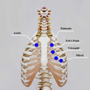
- Follow a systematic pattern when auscultating heart sounds. See Figure 5.18[20] for an illustration of thoracic landmarks for auscultation. The traditional order is as follows:
- Aortic area: Second right intercostal space
- Pulmonic area: Second left intercostal space
- Erb’s point: Third left intercostal space
- Tricuspid area: Fourth left intercostal space at the lower left sternal border
- Mitral area (also referred to as “apical”): Fifth left intercostal space at the midclavicular line
- In each of these areas, listen for the normal heart sounds (S1 and S2). S1 is the first heart sound, often described as “lub,” and corresponds to the closure of the atrioventricular valves (mitral and tricuspid valves). S2 is the second heart sound, described as “dub,” and corresponds to the closure of the semilunar valves (aortic and pulmonic valves). S2 may occur during inspiration, which is considered a normal variation.
- Listen for additional heart sounds, such as S3, S4 murmurs, and pericardial friction rub:
- S3 (also known as a ventricular gallop) is a low frequency heart sound heard shortly after the second heart sound and preceding the normal first heart sound.
- S4 (also known as an atrial gallop) is heard late in the cardiac cycle, just before the first heart sound.
- Murmurs are whooshing or swishing sounds made by rapid, choppy (turbulent) blood flow through the heart that may indicate valvular heart abnormalities. Note the timing, intensity, and location of any murmurs.
- Pericardial friction rub is a high-pitched, scratchy sound that occurs when the inflamed pericardial layers rub against each other. It is typically heard in clients with pericarditis.
Peripheral Assessment
Edema
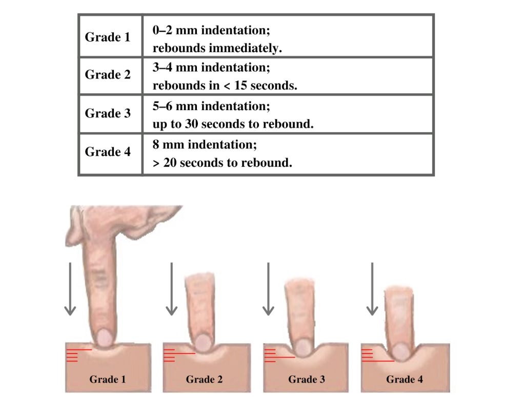
Edema occurs as a result of a buildup of fluid within the tissues. If edema is present on inspection, palpate the area to determine if the edema is pitting or nonpitting. Press on the skin to assess for indentation, ideally over a bony structure, such as the tibia. If no indentation occurs, it is referred to as nonpitting edema. If indentation occurs, it is referred to as pitting edema.
Note the depth of the indention and how long it takes for the skin to rebound back to its original position. The indentation and time required to rebound to the original position are graded on a scale from 1 to 4. Edema rated at 1+ indicates a barely detectable depression with immediate rebound, and 4+ indicates a deep depression with a time lapse of over 20 seconds required to rebound. See Figure 5.19[21] for an illustration of grading edema. Additionally, it is helpful to note edema may be difficult to observe in larger clients. It is also important to monitor for sudden changes in weight, which is considered a probable sign of fluid volume overload.
Capillary Refill
The capillary refill test is performed on the nail beds to monitor perfusion, the amount of blood flow to tissue. Pressure is applied to a fingernail or toenail until it pales, indicating that the blood has been forced from the tissue under the nail. This paleness is called blanching. Once the tissue has blanched, pressure is removed. Capillary refill time is defined as the time it takes for the color to return after pressure is removed. If there is sufficient blood flow to the area, a pink color should return within two to three seconds after the pressure is removed.
Read additional information about performing a focused cardiovascular assessment in the “Cardiovascular Assessment” chapter in Open RN Nursing Skills, 2e.
Life Span Considerations
Nurses consider characteristics of the pediatric clients and older adults during assessment.[22],[23]
Pediatric Considerations
- Many profound changes occur in a newborn’s cardiovascular anatomy and physiology over the first few days and weeks following birth. It is important to distinguish normal fetal to newborn cardiovascular change and signs of abnormality such as congenital heart defects.
- Newborns have incomplete sympathetic innervation and experience greater vulnerability to environmental and physiological influences without the ability to compensate.
- Infants have higher heart rates and lower blood pressures than adults.
- A small volume of blood loss in small children can be significant. For example, 100 mL of blood loss in an infant weighing 5 kg is 10% of their total blood volume.
- Establishing IV access in infants and children can be challenging due to their small veins and increased subcutaneous tissue.
Older Adult Considerations
- Cardiac valves (especially the mitral and aortic valves) often calcify in older adults, impacting their functionality and resulting in murmurs.
- Pacemaker cells decrease in number, increasing the risk of dysrhythmias.
- Increased fibrous tissue and fat in the SA node can impact the conduction of electrical impulses to the atria.
- Increased conduction time occurs (i.e., the amount of time for an electrical impulse to travel through the SA node, atria, the AV node, the bundle of His, the Purkinje fibers, and ventricles).
- The left ventricle increases in size, impacting the quantity of blood and strength of the contractions.
- Decreased sensitivity of baroreceptors and arteriosclerosis can cause orthostatic hypotension.
- Limited fluid intake (due to prescribed fluid restrictions, an attempt to manage incontinence, or decreased mobility in obtaining and drinking fluids) creates viscous blood and increases the risk for developing thrombi.
- Varicosities may develop due to the loss of elasticity of the veins, weaker leg muscles, and muscle atrophy from less exercise.
Laboratory and Diagnostic Testing
Laboratory and diagnostic testing are ordered by health care providers to identify and diagnose cardiovascular conditions.
Laboratory Studies
Several key laboratory values that play a vital role in cardiovascular assessment include troponins, CK (creatine kinase), myoglobin, lipids (total cholesterol, triglycerides, HDL, and LDL), CRP (C-reactive protein), B-type natriuretic peptide, electrolytes, coagulation studies, and thyroid studies. Review normal reference ranges for common diagnostic tests in “Appendix A – Normal Reference Ranges.”
- Troponins: Troponins are myocardial muscle proteins released into the bloodstream with injury to myocardial muscle. Elevated troponin levels indicate cardiac necrosis or an acute myocardial infarction (MI). Troponin T and I are highly specific to cardiac tissue, making them essential markers for diagnosing cardiac events.[24]
- CK (creatine kinase) and CK-MB: CK is an enzyme specific to cells of the brain, myocardium, and skeletal muscle. CK-MB is primarily found in myocardial muscle. CK-MB levels show a predictable rise and fall during three days, with a peak level occurring around 24 hours after the onset of chest pain. Monitoring CK and CK-MB aids in the diagnosis and assessment of MI.[25]
- Myoglobin: Myoglobin is a low-molecular-weight heme protein found in cardiac and skeletal muscle. It is the earliest marker detected after an MI, appearing as early as two hours after the event and declining rapidly after seven hours. However, myoglobin is not cardiac specific, making it less reliable than troponins for MI diagnosis.[26]
- B-type natriuretic peptide (BNP): BNP is a protein that is released by the heart, primarily in response to increased pressure or stress on the heart muscle. This protein plays a crucial role in regulating blood volume and blood pressure. Elevated levels of BNP in the blood can be indicative of heart failure.[27]
- Lipids: Lipid panel laboratory tests include the examination of high-density lipoproteins (HDL), low-density lipoproteins (LDL), and triglycerides. Total cholesterol is a combined measurement of the HDL and LDL numbers. Lipid profile assessments are crucial for identifying cardiovascular risk factors. Elevated total cholesterol, triglycerides, and LDL levels are associated with atherosclerosis and an increased risk of coronary artery disease (CAD). Conversely, higher levels of HDL are considered protective against CAD.[28]
- CRP (C-reactive protein): CRP is the most studied marker of inflammation. Elevated CRP levels can result from any inflammatory process in the body. While not specific to cardiovascular assessment, measuring CRP is valuable for assessing cardiovascular risk, especially in middle-aged or older individuals. Higher CRP levels may indicate an increased risk of cardiovascular events.[29]
- Electrolytes: Many electrolyte imbalances can affect the heart’s electrical activity and muscle function[30]:
- Sodium (Na+): Sodium is the most common electrolyte in extracellular fluid and plays a crucial role in regulating blood pressure, maintaining fluid balance, and transmitting nerve impulses. Elevated sodium levels can cause high blood pressure and edema.
- Potassium (K+): Potassium is the most common electrolyte inside cells and is essential for normal muscle and nerve function, including heart rhythm. Abnormal potassium levels can result in arrhythmias and muscle weakness and can be caused by kidney disease and certain medications.
- Calcium (Ca2+): Calcium is essential for muscle contraction, blood clotting, and bone health. It is tightly regulated by hormones such as the parathyroid hormone and calcitonin. Abnormal calcium levels can affect muscle and nerve function and bone health.
- Magnesium (Mg2+): Magnesium is involved in numerous biochemical processes, including muscle and nerve function, energy metabolism, and bone health. Abnormal magnesium levels can lead to muscle cramps, irregular heart rhythms, and other issues.
- Coagulation studies: Coagulation studies assess the blood’s ability to clot and include prothrombin time (PT), international normalized ratio (INR), and activated partial thromboplastin time (aPTT). Abnormal results can indicate a risk of clot formation or bleeding.
- Prothrombin time (PT): PT measures the time it takes for blood to clot after a substance called thromboplastin is added to a blood sample. It primarily evaluates the extrinsic and common coagulation pathways. PT is often used to monitor the effect of anticoagulant medications like warfarin and to diagnose bleeding disorders.
- International normalized ratio (INR): INR is a standardized measurement derived from the PT. It is used to ensure consistency in PT results between different laboratories. INR is particularly important for individuals on anticoagulant therapy (e.g., warfarin) to monitor and maintain their blood’s clotting ability within a target range.
- Activated partial thromboplastin time (aPTT): aPTT measures the time it takes for blood to clot after a substance that activates the intrinsic and common coagulation pathways is added to a blood sample. It is used to assess factors involved in the clotting cascade, including factors VIII, IX, XI, and XII. aPTT is used to diagnose and monitor conditions like hemophilia and to evaluate the effects of anticoagulant medications such as heparin.
- Thrombin time (TT): TT measures the time it takes for fibrinogen to convert into fibrin. It primarily assesses the final step of the clotting process. TT may be used to evaluate specific clotting disorders and fibrinogen abnormalities.
- D-dimer: D-dimer is a marker for the presence of blood clots, specifically the breakdown products of fibrin that result from the dissolution of a blood clot. Elevated levels of D-dimer may suggest the presence of an active blood clotting process in the body. D-dimer tests are often used in the diagnosis of conditions like deep vein thrombosis (DVT), pulmonary embolism (PE), and disseminated intravascular coagulation (DIC).[31]
- Thyroid function tests: Thyroid hormones help regulate heart rate and cardiac output. Hypothyroidism can lead to bradycardia (slower heart rate), increased cholesterol levels, and increased risk of atherosclerosis, which can contribute to cardiovascular disease. Conversely, hyperthyroidism can cause tachycardia (rapid heart rate) and increased cardiac output, potentially leading to conditions like atrial fibrillation or heart failure. Thyroid hormones also play an important role in regulating the sensitivity of the heart to adrenaline (epinephrine) and norepinephrine, which are hormones that influence heart rate and contractility. Abnormal levels of T4 and T3 can disrupt these processes and affect heart function.[32]
Diagnostic Testing
Providers who suspect a cardiac disorder may initially order a chest X-ray to assess the size of cardiac structures and to check for fluid in the lungs. Other common diagnostic tests related to the cardiovascular system include electrocardiograms (ECG), Holter monitors, echocardiograms, cardiac stress tests, and cardiac catheterization.
Electrocardiogram
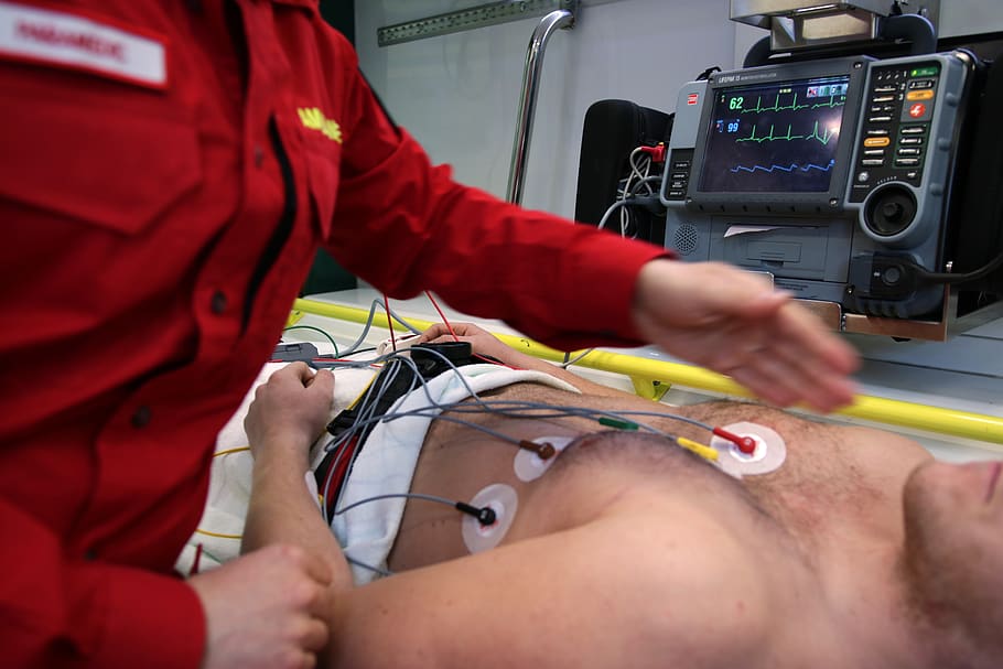
An electrocardiogram (EKG or ECG) is an important diagnostic tool to help identify cardiac abnormalities. See Figure 5.20[33] for an image of a client undergoing an ECG and Figure 5.21[34] for an image of a normal ECG strip showing normal sinus rhythm. ECG records the electrical activity of the heart and allows health care professionals to assess the heart’s rhythm and identify various cardiac conduction abnormalities called dysrhythmias or arrhythmias. Cardiac dysrhythmias can range from asymptomatic conditions to life-threatening events that impair the delivery of oxygenated blood to tissues and organs. See Table 5.3b for a summary of common dysrhythmias. An ECG is also a critical tool for diagnosing myocardial ischemia (i.e., a “heart attack”). Specific changes in an ECG tracing can reflect elevation or depression of the ST segment, allowing a provider to identify where potential ischemia or infarction of cardiac tissue may be occurring within the heart.[35]

Nurses must recognize abnormal ECG rhythms requiring prompt intervention. View images of ECGs for Abnormal Rhythms in Nursing Advanced Skills, Chapter 7 Appendix.
Table 5.3b. Common Cardiac Arrhythmias
| Cardiac Rhythm | Rate | Significance |
|---|---|---|
| Normal Sinus Rhythm | 60-100 beats per minute (bpm) | Healthy, regular heart rhythm. |
| Sinus Bradycardia | Fewer than 60 bpm in adults | May be normal for well-conditioned athlete, a side effect of medications, or a symptom of various medical conditions. May require a pacemaker if the client is symptomatic. |
| Sinus Tachycardia | Greater than 100 bpm in adults | May be caused by fever, stress, pain, or hypoxia or a symptom of various medical conditions. Transient sinus tachycardia is a normal finding during strenuous physical activity. |
| Atrial Fibrillation (A-Fib) | Irregularly irregular rhythm and rate | Characterized by atrial quivering that can cause decreased cardiac output because the ventricles are not able to fill and pump the appropriate amount of blood with each beat. A-fib increases the risk of stroke. May require a pacemaker. A-fib is a common cardiac arrhythmia with aging individuals. |
| Atrial Flutter | Regular rate | Atrial impulses are fast and regular, with rates between 250-300, and the ventricular rate is not the same as the atrial rate. As a result, the client’s cardiac output decreases. |
| Ventricular Tachycardia (VT or V-tach) | Greater than 100 beats per minute | The ventricular rate is often over 120 beats per minute, resulting in rapidly worsening cardiac output. The client may only be able to tolerate this rapid ventricular rhythm for a short period of time before losing consciousness. V-tach requires emergency response. |
| Ventricular Fibrillation (VF) | No heart rate | Characterized by quivering ventricles with no effective contractions and no cardiac output. This is the most dangerous arrhythmia because of lack of cardiac output and requires immediate initiation of CPR and emergency response. |
| First-Degree Heart Block | Variable | Characterized by a prolonged interval between atrial and ventricular contraction. Can be normal or an early sign of degeneration that can progress to more severe blocks. |
| Second-Degree Heart Block | Variable | Characterized by dropped ventricular beats that can result in decreased cardiac output. May require a pacemaker if symptomatic. |
| Third-Degree Heart Block | Variable | Characterized by a complete electrical dissociation between the atria and ventricles. May require a pacemaker to help maintain adequate cardiac output. |
Read additional information about abnormal heart rhythms and view ECG strips in the “Interpret Basic ECG” chapter in Open RN Nursing Advanced Skills.
Pacemakers
Pacemakers may be inserted in clients to restore normal cardiac conduction and minimize the impact of arrythmias on cardiac output. Pacemakers send electrical impulses to mimic the coordinated actions of the sinus to AV node conduction, restoring efficiency and synchronization to the contraction and relaxation of the heart tissue. Pacemakers are a relatively small battery-powered device that has leads connected to heart tissue. The device can sense the inherent electrical impulses of the heart tissue and override them when needed. The pacemaker includes a generator, a battery, and a small computer to regulate the heart impulses. The pacemaker leads include insulated wires that can be placed inside or onto the cardiac tissue. Pacemakers may be temporary or permanent, depending on the underlying cause of the cardiac arrythmia.
- Transvenous pacemakers are inserted through a vein and threaded into the heart. The generator is commonly placed in a small pectoral pocket in the upper chest.
- Epicardial pacemakers are inserted through an incision in the chest and the leads are attached to the heart. The pacemaker is inserted into a pocket in the skin of the abdomen.
A cardiac provider will determine the type of pacemaker that is required and the settings that will restore appropriate cardiac function to the client. A single chamber pacemaker sends impulses to the right ventricle, whereas a dual-chamber pacemaker can send signals to the upper and lower right heart chambers. Biventricular pacemakers stimulate both lower ventricles of the heart and are used in situations of advanced heart failure.
People who have a permanent pacemaker will require periodic monitoring. The status of the pacemaker will be regularly checked or “interrogated” (often done remotely using a telephone or a secure web-based system) to provide information regarding the heart rhythm, the functioning of the pacemaker leads/generator, the electrical activity initiated by the pacemaker, or inherent to the heart, the battery life and the presence of any abnormal heart rhythms. Individuals with implantable pacemakers should be cautioned regarding the use of magnetic devices directly over the device and should never undergo MRI imaging.[36]
Holter Monitors
Holter monitors are portable devices that continuously record a client’s heart rhythm and electrical activity over an extended period, typically 24 to 48 hours. See Figure 5.22[37] for an illustration of a Holter monitor. Its primary purpose is to detect and document irregularities in the heart’s electrical patterns, such as arrhythmias, which may not be captured during brief in-office ECGs. Additionally, clients may activate a trigger on a Holter device to denote the occurrence of cardiac symptoms, such as increased shortness of breath, fatigue, or dizziness. A cardiologist can then review the symptom timestamp and accompanying EGC tracing to identify if an arrhythmia occurred at that time.[38]
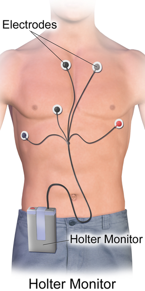
By providing a thorough real-time analysis of a client’s heart rate and rhythm during their daily activities, the results of a Holter monitor help health care professionals identify and diagnose a variety of cardiac conditions, including atrial fibrillation, ventricular tachycardias, bradycardias, and heart blocks.
Echocardiogram
A transthoracic echocardiogram is a noninvasive diagnostic test that uses sound waves (ultrasound) to create real-time images of the heart’s structure and function. See Figure 5.23[39] for an image of a pediatric client undergoing a transthoracic echocardiogram. A trained sonographer or cardiologist applies a gel and a transducer to the client’s chest. The transducer is moved to different areas of the chest to obtain various views of the heart, including the heart chambers, valves, walls, and blood flow patterns. Doppler ultrasound is also commonly used during echocardiography to determine the speed and direction of blood flow through the valves and chambers of the heart.
A transesophageal echocardiogram (TEE) is similar to transthoracic although it is invasive in nature. The provider may order a transesophageal echocardiogram to gain a more detailed view of the heart and aorta. To perform a transesophageal echocardiogram, a probe and ultrasound wand are guided down the esophagus and close to the position of the heart, and the ultrasound is performed. Conscious sedation is administered to enhance client comfort. Both diagnostic tools are very helpful for identifying heart failure, valvular regurgitation, and stenosis.[40],[41]
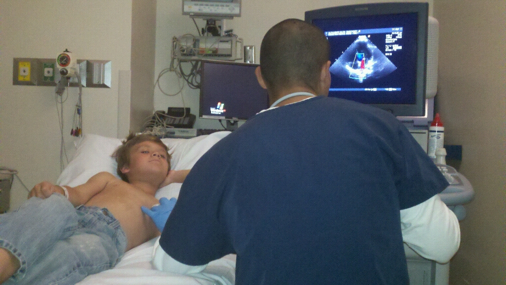
A key measurement during an echocardiogram is the client’s ejection fraction. Ejection fraction measures the amount of blood the left ventricle of the heart pumps out to the body with each heartbeat. For example, an ejection fraction of 60 percent means that 60 percent of the total amount of blood in the left ventricle is pushed out with each heartbeat. A normal heart’s ejection fraction is between 55 and 70 percent. The ejection fraction is used to diagnose and monitor heart failure.
Cardiac Stress Test
A cardiac stress test (also known as an exercise stress test or treadmill test) is a diagnostic procedure used to evaluate the performance and function of the heart during physical activity. It helps identify potential heart problems, such as coronary artery disease (CAD), arrhythmias, and heart valve issues. During a cardiac stress test procedure, a client is asked to walk on a treadmill or pedal a stationary bicycle with the exercise requirement gradually increasing over time. See Figure 5.24[42] for an image of a cardiac stress test.
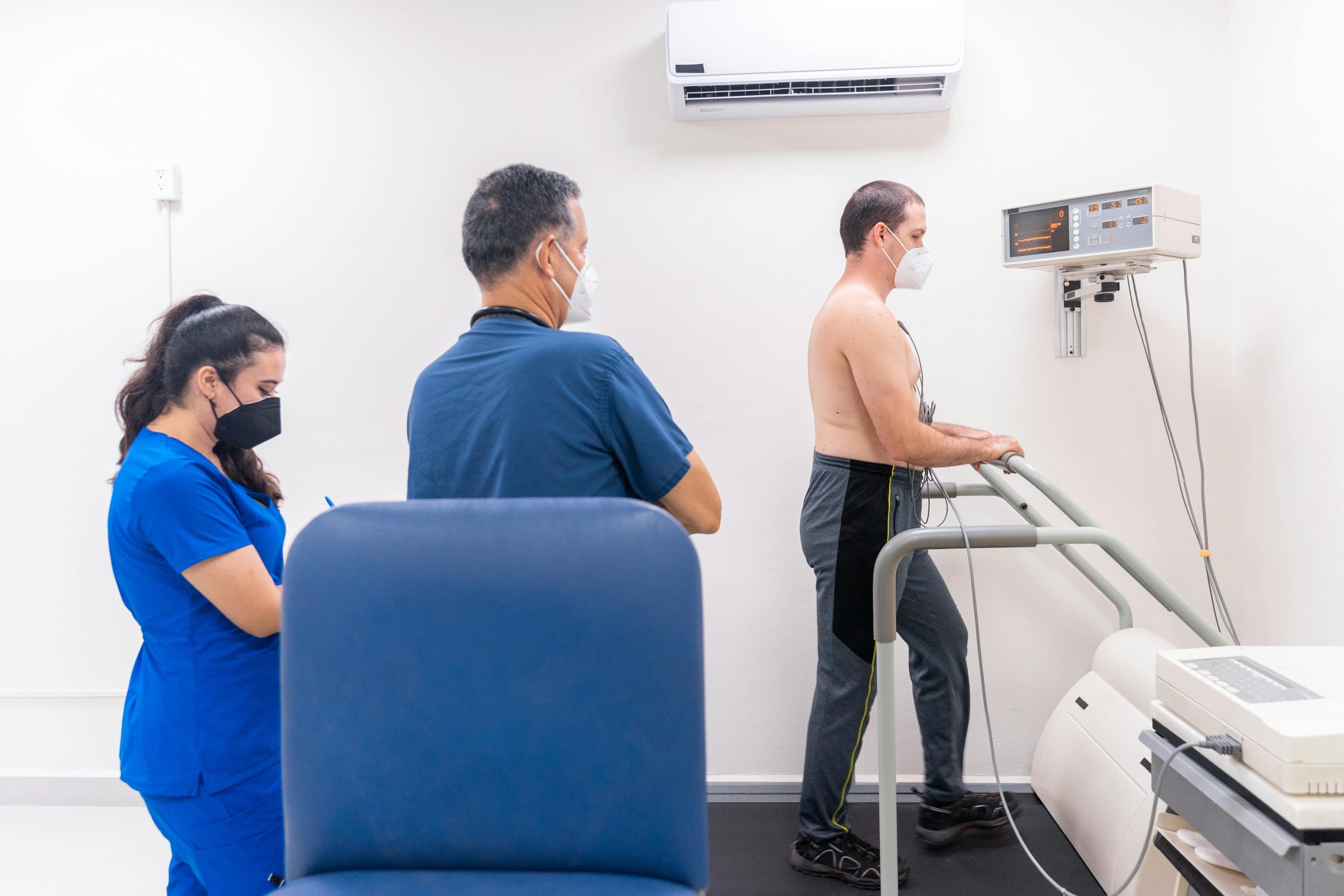
During the procedure, client symptoms are monitored for occurrence of chest pain, shortness of breath, fatigue, and dizziness. The client’s ECG is also closely monitored to detect any changes in heart’s electrical activity during exercise. The test is terminated when the client has achieved a target heart rate, if symptoms of discomfort occur, if the client’s experiences significant ECG changes, or if the maximum duration of exercise has occurred. Cardiac stress tests may also be performed with the administration of medications such as dobutamine or adenosine for individuals who are unable to tolerate exercise-induced stress testing. The medications can simulate the workload that would typically occur during physical exercise.[43]
Cardiac Catheterization
Cardiac catheterization is a valuable diagnostic procedure in the evaluation and management of cardiovascular health alterations. Cardiac catheterization, performed in association with a coronary angiogram, is a diagnostic procedure used to visualize the coronary arteries, heart chambers, and great vessels. It involves the insertion of a thin, flexible tube (catheter) into the blood vessels, usually through the femoral or radial artery, to access the heart and its surrounding structures.[44],[45] See Figure 5.25[46] for an image of a cardiac catheterization procedure with the image of the client’s coronary arteries and heart chambers displayed on the monitor.

Providers order cardiac catheterization for many diagnostic purposes, such as the following:
- Assessment of Coronary Artery Disease (CAD): Identifies blocked or narrowed coronary arteries, guiding decisions about interventions such as angioplasty (use of a balloon to stretch open a blocked artery) or stent (insertion of a small mesh tube to help expand an artery) placement.
- Evaluation of Valvular Heart Disease: Measures pressure gradients across heart valves and assesses valve function.
- Investigation of Congenital Heart Abnormalities: Assesses structural abnormalities of the heart and great vessels in pediatric and adult clients.
- Measurement of Cardiac Function: Directly measures cardiac output, ejection fraction, and other important parameters of cardiac function.
See the following box regarding nursing interventions to prepare a client for cardiac catheterization.
Client Preparation for Cardiac Catheterization[47]
Before a cardiac catheterization procedure, thorough client preparation is needed. This involves collaboration from the nurse and health care team to ensure that all client safety procedural tasks have been addressed.
Informed Consent: The nurse ensures signed informed consent has been obtained and the client understands the procedure, its risks, and alternatives. Concerns are forwarded to the physician before the procedure begins.
Baseline Vital Signs: Baseline vital signs, including blood pressure, heart rate, respiratory rate, and temperature are documented. Monitoring trends in these parameters during the procedure is vital for detecting potential complications.
History/Allergy Assessment: A detailed history, including any known allergies, particularly iodine allergies or previous adverse reactions to contrast agents, is obtained. It is crucial to notify the health care team of a history of iodine or iodinated contrast media allergy because contrast agents are commonly used during cardiac catheterization. Allergic reactions can range from mild rashes to severe anaphylactic reactions. Clients with known allergies may require alternative contrast media or premedication to reduce the risk of an allergic response. The client’s renal function should also be assessed because contrast dye can impact renal function.
NPO Status: The client must remain NPO (nothing by mouth) for a specified period before the procedure (usually 6-8 hours), as dictated by agency guidelines.
Medication Management: The client’s current medications should be reviewed for anticoagulants and antiplatelet agents that pose bleeding risks.
IV Access: Intravenous (IV) access is established for administering medications, contrast, and fluids during the procedure, which may be obtained via peripheral and/or central access.
Groin Prep: The groin area is prepared because the femoral artery is often used as the access point for inserting catheters. Prep involves shaving or clipping hair according to agency guidelines and cleansing the area with an antiseptic solution, such as chlorhexidine. If a radial approach is used, cleansing of the wrist and radial artery site with antiseptic is sufficient.
Health Teaching and Support: Health teaching is provided, including what to expect during and after the procedure. Emotional support is provided because many clients experience anxiety related to this invasive procedure.
During cardiac catheterization, a catheter is inserted through the access site and advanced through the blood vessels until it reaches the coronary arteries or the heart chambers. Contrast dye is injected through the catheter, and X-ray imaging is used to visualize the coronary arteries and identify areas of narrowing or blockage. Measurements of pressures and blood oxygen saturations within the heart may be taken to identify abnormalities in valve function. If areas of blockage or narrowing are noted within the coronary arteries, an angioplasty may be performed where a balloon is inflated to help open the coronary artery. After the balloon is inflated, a stent is often inserted to help keep the coronary artery open. After the procedure has been completed, the catheter is removed, and pressure is applied to the access site until bleeding stops.[48] Clients will receive instruction to minimize movement and pressure to the catheter insertion site for a specified period. The site should be closely monitored for recurrent bleeding and pressure should be immediately applied to the insertion site if bleeding recurs. Urinary output is also closely monitored to ensure the contrast dye has not impeded renal function. Additional post-procedure education should include assessing for infection, reporting any signs of chest discomfort, knowing the guidelines for activity progression, etc.
See the following box for a case study related to a cardiac catheterization procedure.
Cardiac Catheterization Case Study
Michael is a 58-year-old man who has been experiencing recurrent episodes of chest pain and shortness of breath. His medical history includes hypertension, hyperlipidemia, and a family history of coronary artery disease (CAD). After a thorough evaluation, it has been determined that a coronary angiography (heart catheterization) is necessary to assess the extent of his coronary artery disease.
1. What are the key steps and considerations in preparing Michael for a coronary angiography procedure?
Medical history, allergies, current medications, renal function, baseline vitals/assessment, client comfort, IV access.
2. How can health care providers ensure that Michael provides informed consent for the coronary angiography procedure?
Provide verbal instruction/written materials or brochures explaining coronary angiography, its purpose, benefits, risks, alternate procedures, and expectations before, during, and after the procedure.
3. Following the coronary angiography, what immediate post-procedure monitoring should be in place for Michael?
Vital signs monitoring, cardiac monitoring, access site monitoring, fluid balance, urine output.
Media Attributions
- pexels-photo-4262171
- Cardiac Ausculation image8-768×768
- Grading Edema
- ambulance-emergency-medic-health-vehicle-rescue
- Holter_Monitor
- 5619843496_1201a6a160_o
- A_cardiac_catheterization_procedure_in_the_Naval_Medical_Center_San_Diego_hospital’s_cardiac_catheterization_laboratory
- National Heart, Lung, and Blood Institute. (2022, May 18). What Is metabolic syndrome? National Institutes of Health. https://www.nhlbi.nih.gov/health/metabolic-syndrome ↵
- Wilson, P. (2023). Overview of established risk factors for cardiovascular disease. UpToDate. Retrieved August 23, 2023, from https://www.uptodate.com/ ↵
- “food1.jpg” by bigbrand . is licensed under CC BY 2.0 ↵
- Fawzy, A. M., & Lip, G. Y. H. (2021). Cardiovascular disease prevention: Risk factor modification at the heart of the matter. The Lancet Regional Health Western Pacific, 17, 100291. https://pubmed.ncbi.nlm.nih.gov/34734203/ ↵
- American Heart Association. (2022, December 6). Understand your risks to prevent a heart attack. https://www.heart.org/en/health-topics/heart-attack/understand-your-risks-to-prevent-a-heart-attack ↵
- Wilson, P. (2023). Overview of established risk factors for cardiovascular disease. UpToDate. Retrieved August 23, 2023, from https://www.uptodate.com/ ↵
- “family-setting-the-table-for-dinner” by August de Richlieu, via Pexels.com is licensed under CC0 ↵
- Boudi, F. B. (2021, October 21). Risk factors for coronary artery disease. Medscape. https://emedicine.medscape.com/article/164163-overview ↵
- Schultz, W. M., Heval, K., Lisko, J., Varghese, T., Shen, J., Sandesara, P., Quyyumi, A. A., Taylor, H., Gulati, M., Harold, J., Mieres, J., Ferdinand, K., Menash, G., & Sperling, L. (2018). Socioeconomic status and cardiovascular outcomes. Circulation, 137(20), 2166-2178. https://doi.org/10.1161/circulationaha.117.029652 ↵
- Schultz, W. M., Heval, K., Lisko, J., Varghese, T., Shen, J., Sandesara, P., Quyyumi, A. A., Taylor, H., Gulati, M., Harold, J., Mieres, J., Ferdinand, K., Menash, G., & Sperling, L. (2018). Socioeconomic status and cardiovascular outcomes. Circulation, 137(20), 2166-2178. https://doi.org/10.1161/circulationaha.117.029652 ↵
- Schultz, W. M., Heval, K., Lisko, J., Varghese, T., Shen, J., Sandesara, P., Quyyumi, A. A., Taylor, H., Gulati, M., Harold, J., Mieres, J., Ferdinand, K., Menash, G., & Sperling, L. (2018). Socioeconomic status and cardiovascular outcomes. Circulation, 137(20), 2166-2178. https://doi.org/10.1161/circulationaha.117.029652 ↵
- Centers for Disease Control and Prevention. (2023, May 15). About heart disease. https://www.cdc.gov/heartdisease/about.htm ↵
- Colucci, W. S., & Borlaug, B. A. (2022). Heart failure: Clinical manifestations and diagnosis in adults. UpToDate. Retrieved August 28, 2023, from https://www.uptodate.com/ ↵
- Basile, J., & Bloch, M. J. (2023). Overview of hypertension in adults. UpToDate. Retrieved August 29, 2023, from https://www.uptodate.com/ ↵
- Crea, F., Kolodgie, F., Finn, A. & Virmani, R. (2022). Mechanisms of acute coronary syndromes related to atherosclerosis. UpToDate. Retrieved August 20, 2023, from https://www.uptodate.com/ ↵
- Neschis, D. G., & Golden, M. A. (2022). Clinical features and diagnosis of lower extremity peripheral artery disease. UpToDate. Retrieved August 30, 2023, from https://www.uptodate.com/ ↵
- Dalman, R. L., & Mell, M. (2023). Overview of abdominal aortic aneurysm. UpToDate. Retrieved September 1, 2023, from https://www.uptodate.com/ ↵
- Spelman, D. (2022). Complications and outcome of infective endocarditis. UpToDate. Retrieved September 2, 2023, from https://www.uptodate.com/ ↵
- Meyer, T. E. (2022). Auscultation of heart sound. UpToDate. Retrieved August 24, 2023, from https://www.uptodate.com/ ↵
- “Cardiac Auscultation Areas” by Meredith Pomietlo for Chippewa Valley Technical College is licensed under CC BY 4.0 ↵
- “Grading of Edema” by Meredith Pomietlo for Chippewa Valley Technical College is licensed under CC BY 4.0 ↵
- Queensland Pediatric Emergency Care. (2022). How children are different - Anatomical and physiological differences. https://www.childrens.health.qld.gov.au/__data/assets/pdf_file/0031/179725/how-children-are-different-anatomical-and-physiological-differences.pdf ↵
- Saikia, D., & Mahanta, B. (2019). Cardiovascular and respiratory physiology in children. Indian Journal of Anaesthesia, 63(9), 690–697. https://doi.org/10.4103/ija.IJA_490_19. ↵
- Mayo Clinic. (2022, January 15). Blood tests for heart disease. https://www.mayoclinic.org/diseases-conditions/heart-disease/in-depth/heart-disease/art-20049357 ↵
- Mayo Clinic. (2022, January 15). Blood tests for heart disease. https://www.mayoclinic.org/diseases-conditions/heart-disease/in-depth/heart-disease/art-20049357 ↵
- Mayo Clinic. (2022, January 15). Blood tests for heart disease. https://www.mayoclinic.org/diseases-conditions/heart-disease/in-depth/heart-disease/art-20049357 ↵
- Mayo Clinic. (2022, January 15). Blood tests for heart disease. https://www.mayoclinic.org/diseases-conditions/heart-disease/in-depth/heart-disease/art-20049357 ↵
- Mayo Clinic. (2022, January 15). Blood tests for heart disease. https://www.mayoclinic.org/diseases-conditions/heart-disease/in-depth/heart-disease/art-20049357 ↵
- Mayo Clinic. (2022, January 15). Blood tests for heart disease. https://www.mayoclinic.org/diseases-conditions/heart-disease/in-depth/heart-disease/art-20049357 ↵
- Mayo Clinic. (2022, January 15). Blood tests for heart disease. https://www.mayoclinic.org/diseases-conditions/heart-disease/in-depth/heart-disease/art-20049357 ↵
- Mayo Clinic. (2022, January 15). Blood tests for heart disease. https://www.mayoclinic.org/diseases-conditions/heart-disease/in-depth/heart-disease/art-20049357 ↵
- Mayo Clinic. (2022, January 15). Blood tests for heart disease. https://www.mayoclinic.org/diseases-conditions/heart-disease/in-depth/heart-disease/art-20049357 ↵
- “ambulance-emergency-medic-health-vehicle-rescue.jpg” by unknown author, via pxfuel.com is licensed under CC0 ↵
- “Normal Sinus Rhythm” by Deanna Hoyord is licensed under CC BY 4.0 ↵
- Prutkin, J. M. (2023). ECG tutorial: Basic principles of ECG analysis. UpToDate. Retrieved August 25, 2023, from https://www.uptodate.com/ ↵
- Olshansky, B. (2022, Nov.). Patient education: Pacemakers (Beyond the basics). UpToDate. Retrieved August 25, 2023, from https://www.uptodate.com/contents/pacemakers-beyond-the-basics ↵
- “Holter_Monitor.png” by BruceBlaus is licensed under CC BY-SA 4.0 ↵
- Madias, C. (2022). Ambulatory ECG monitoring. UpToDate. Retrieved August 25, 2023, from https://www.uptodate.com/ ↵
- “5619843496” by Jeremy Miles is licensed under CC BY-SA 2.0 ↵
- Johns Hopkins Medicine. (n.d.). Echocardiogram. https://www.hopkinsmedicine.org/health/treatment-tests-and-therapies/echocardiogram ↵
- Mayo Clinic. (n.d.). Echocardiogram. https://www.mayoclinic.org/tests-procedures/echocardiogram/about/pac-20393856 ↵
- “a-cardiologist-examining-a-patient-undergoing-cardiac-stress-test” by Los Muertos Crew, via Pexels.com is licensed under CC0 ↵
- Askew, J. W., Chareonthaitawee, P., & Arruda-Olsen, A. M. (2022). Selecting the optimal cardiac stress test. UpToDate. Retrieved August 26, 2023, from https://www.uptodate.com/ ↵
- Kern, M. J. (2022). Cardiac catheterization techniques: Normal hemodynamics. UpToDate. Retrieved August 26, 2023, from https://www.uptodate.com/ ↵
- Velagapudi, P. (2022). Preparing patients for cardiac catheterization and possible coronary artery intervention. UpToDate. Retrieved August 27, 2023, from https://www.uptodate.com/ ↵
- “A_cardiac_catheterization_procedure_in_the_Naval_Medical_Center_San_Diego_hospital’s_cardiac_catheterization_laboratory.jpg” by Navy Medicine is licensed in the Public Domain. ↵
- Velagapudi, P. (2022). Preparing patients for cardiac catheterization and possible coronary artery intervention. UpToDate. Retrieved August 27, 2023, from https://www.uptodate.com/ ↵
- Kern, M. J. (2022). Cardiac catheterization techniques: Normal hemodynamics. UpToDate. Retrieved August 26, 2023, from https://www.uptodate.com/ ↵
Answer Key to Chapter 5 Learning Activities
Section 5.3 Military Time
- 7:30 PM
- 12:30 AM
- 0900
- 2200
Section 5.4 Household and Metric Equivalents
- 5 mL
- 30 mL
- 500 mg
- 3636 grams
- 0.7 cm
Section 5.5 Rounding
- 6.5
- 6.5
- 5.5
- 5.5
- 0.19
- 0.19
- 0.2
- 0.2
Answers to interactive elements are given within the interactive element.
Use this checklist to perform a "General Survey." Checklists for hand washing, using hand sanitizer, and obtaining vital signs are included in Appendix A.
Steps
Disclaimer: Always review and follow agency policy regarding this specific skill.
- Knock, enter the room, greet the patient, and provide for privacy.
- Introduce yourself, your role, the purpose of your visit, and an estimate of the time it will take.
- Perform hand hygiene.
- Ask the patient their legal name and date of birth to establish two unique identifiers. Verify the information provided in their chart or wristband, if present. Use one of the following for the second verification:
- Scan wristband
- Compare name/DOB to MAR
- Ask staff to verify patient (in settings where wristbands are not worn)
- Compare picture on MAR to patient
- Address patient needs (pain, toileting, glasses/hearing aids) prior to starting assessment. Note if the patient has signs of distress such as difficulty breathing or chest pain. If signs are present, defer general survey and obtain emergency assistance per agency policy.
- Explain the procedure to the patient; ask if they have any questions. Obtain an interpreter as needed if English is not the patient’s primary language.
- Pause and explain to the instructor what you would purposefully observe and assess during a general survey assessment.
- Upon completion of the survey, thank the patient and ask if anything is needed.
- Ensure safety measures when leaving the room:
- CALL LIGHT: Within reach
- BED: Low and locked (in lowest position and brakes on)
- SIDE RAILS: Secured
- TABLE: Within reach
- ROOM: Risk-free for falls (scan room and clear any obstacles)
- Perform hand hygiene and clean stethoscope.
- Follow agency policy for reporting findings outside of normal range.
- Document the assessment.
Learning Activities
(Answers to "Learning Activities" can be found in the "Answer Key" at the end of the book. Answers to interactive activity elements will be provided within the element as immediate feedback.)
Maria is working on a medical surgical unit and receives a direct admission from the internal medicine clinic. She arrives at the patient’s room to complete the initial admission assessment. All of the following conditions are found. Of these conditions, which of the following should be reported immediately to the health care provider.
- Patient ambulates with assistance of wheeled walker.
- Patient’s BMI is outside of the normal range.
- Patient appears unkempt and has strong body odor.
- Patient is experiencing increased difficulty breathing.
"Vital Signs Case Study” by Susan Jepsen for Lansing Community College is licensed under CC BY 4.0
![]()
Test your clinical judgment with an NCLEX Next Generation-style question: Chapter 1, Assignment 1.
![]()
Test your clinical judgment with an NCLEX Next Generation-style question: Chapter 1, Assignment 2.
![]()
Test your clinical judgment with an NCLEX Next Generation-style question: Chapter 1, Assignment 3.
![]()
Test your clinical judgment with an NCLEX Next Generation-style question: Chapter 1, Assignment 4.
![]()
Test your clinical judgment with an NCLEX Next Generation-style question: Chapter 1, Assignment 5.
![]()
Test your clinical judgment with an NCLEX Next Generation-style question: Chapter 1, Assignment 6.
![]()
Test your clinical judgment with an NCLEX Next Generation-style question: Chapter 1, Assignment 7.
Learning Activities
(Answers to "Learning Activities" can be found in the "Answer Key" at the end of the book. Answers to interactive activity elements will be provided within the element as immediate feedback.)
Maria is working on a medical surgical unit and receives a direct admission from the internal medicine clinic. She arrives at the patient’s room to complete the initial admission assessment. All of the following conditions are found. Of these conditions, which of the following should be reported immediately to the health care provider.
- Patient ambulates with assistance of wheeled walker.
- Patient’s BMI is outside of the normal range.
- Patient appears unkempt and has strong body odor.
- Patient is experiencing increased difficulty breathing.
"Vital Signs Case Study” by Susan Jepsen for Lansing Community College is licensed under CC BY 4.0
Test your clinical judgment with an NCLEX Next Generation-style question: Chapter 1, Assignment 1.
Test your clinical judgment with an NCLEX Next Generation-style question: Chapter 1, Assignment 2.
Test your clinical judgment with an NCLEX Next Generation-style question: Chapter 1, Assignment 3.
Test your clinical judgment with an NCLEX Next Generation-style question: Chapter 1, Assignment 4.
Test your clinical judgment with an NCLEX Next Generation-style question: Chapter 1, Assignment 5.
Test your clinical judgment with an NCLEX Next Generation-style question: Chapter 1, Assignment 6.
Test your clinical judgment with an NCLEX Next Generation-style question: Chapter 1, Assignment 7.
Sample Documentation of Expected Findings
Mrs. Smith is a 65-year-old patient who appears her stated age. Calm, cooperative, alert, and oriented x 3. Well-groomed with clean clothing and appropriate for weather. Speech is clear, understandable, and follows instructions appropriately. Moves all extremities equally bilaterally with good posture. Gait is smooth and maintains balance without assistance. Skin warm and mucous membranes moist. 5’4” and weighs 143 pounds with BMI of 24 in normal weight category. Vital signs: BP 120/70, pulse 74 and regular, respiratory rate 14, temperature 36.8 Celsius, SpO2 98% on room air.
Sample Documentation of Unexpected Findings
Mrs. Smith is a 65-year-old patient with older appearance than stated age. Slightly agitated during the interview. Oriented to person only and denies pain. Wearing a heavy winter coat on a warm summer day and unclean body odor. Slow to respond to questions and does not follow commands. Neglect noted of right arm. Gait shuffling with stooped posture with no assistive device. 5’4” and weighs 102 pounds with BMI of 17.5 in the underweight category. Vital signs: BP 186/55, pulse 102 and irregular, respiratory rate 22, temperature 38.1 Celsius, and SpO2 88% on room air.
Affect: Outward display of one’s emotional state. A “flat” affect with little display of emotion is associated with depression.
AIDET: Mnemonic for introducing oneself in health care that includes Acknowledge, Introduce, Duration, Explanation, and Thank You.[1]
Auscultation: Listening to sounds, such as heart, lung, and bowel sounds, created by organs using a stethoscope.
BMI: A standardized reference range to gauge a patient’s weight status.
Cultural safety: The creation of safe spaces for patients to interact with health professionals without judgment, racial reductionism, racialization, or discrimination.
Developmental stages: A person’s life span can be classified into nine categories of development, including Prenatal Development, Infancy and Toddlerhood, Early Childhood, Middle Childhood, Adolescence, Early Adulthood, Middle Adulthood, Late Adulthood, and Death and Dying.
Family dynamics: Patterns of interactions between family members that influence family structure, hierarchy, roles, values, and behaviors.
General survey assessment: A component of a patient assessment that observes the entire patient as a whole. Observation includes using all five senses to gather cues that provide a guideline for additional focused assessments in areas of concern.
Inspection: The observation of a patient’s anatomical structures.
Medical asepsis: Measures to prevent the spread of infection in health care agencies.
Objective data: Information observed through your sense of hearing, sight, smell, and touch while assessing the patient.
Older adults: People over the age of 65.
Percussion: An advanced physical examination technique where body parts are tapped with fingers to determine their size and if fluid is present.
Personal Protective Equipment (PPE): Includes gloves, gowns, goggles, face shields, and masks, along with environmental controls, to prevent the transmission of infection for patients who are diagnosed or suspected of having an infectious disease.
Physical examination: A systematic data collection method of the body that uses the techniques of inspection, auscultation, palpation, and percussion.
Primary data: Information provided directly by the patient.
Primary survey: A brief observation at the start of a shift or visit to verify the patient is stable by assessing mental status, airway, breathing, and circulation.
Secondary data: Information collected from a family member, chart, or other sources.
Subjective data: Information obtained from the patient and/or family members that offers important cues from their perspectives.
Affect: Outward display of one’s emotional state. A “flat” affect with little display of emotion is associated with depression.
AIDET: Mnemonic for introducing oneself in health care that includes Acknowledge, Introduce, Duration, Explanation, and Thank You.[2]
Auscultation: Listening to sounds, such as heart, lung, and bowel sounds, created by organs using a stethoscope.
BMI: A standardized reference range to gauge a patient’s weight status.
Cultural safety: The creation of safe spaces for patients to interact with health professionals without judgment, racial reductionism, racialization, or discrimination.
Developmental stages: A person’s life span can be classified into nine categories of development, including Prenatal Development, Infancy and Toddlerhood, Early Childhood, Middle Childhood, Adolescence, Early Adulthood, Middle Adulthood, Late Adulthood, and Death and Dying.
Family dynamics: Patterns of interactions between family members that influence family structure, hierarchy, roles, values, and behaviors.
General survey assessment: A component of a patient assessment that observes the entire patient as a whole. Observation includes using all five senses to gather cues that provide a guideline for additional focused assessments in areas of concern.
Inspection: The observation of a patient’s anatomical structures.
Medical asepsis: Measures to prevent the spread of infection in health care agencies.
Objective data: Information observed through your sense of hearing, sight, smell, and touch while assessing the patient.
Older adults: People over the age of 65.
Percussion: An advanced physical examination technique where body parts are tapped with fingers to determine their size and if fluid is present.
Personal Protective Equipment (PPE): Includes gloves, gowns, goggles, face shields, and masks, along with environmental controls, to prevent the transmission of infection for patients who are diagnosed or suspected of having an infectious disease.
Physical examination: A systematic data collection method of the body that uses the techniques of inspection, auscultation, palpation, and percussion.
Primary data: Information provided directly by the patient.
Primary survey: A brief observation at the start of a shift or visit to verify the patient is stable by assessing mental status, airway, breathing, and circulation.
Secondary data: Information collected from a family member, chart, or other sources.
Subjective data: Information obtained from the patient and/or family members that offers important cues from their perspectives.
"'Sickness' is what is happening to the patient. Listen to them."[3]
The profession of nursing is defined by the American Nurses Association as “the art and science of caring and focuses on the protection, promotion, and optimization of health and human functioning; prevention of illness and injury; facilitation of healing; and alleviation of suffering through compassionate presence. Nursing is the diagnosis and treatment of human responses and advocacy in the care of individuals, families, groups, communities, and populations in recognition of the connection of all humanity.[4] Simply put, nurses treat human responses to health problems and/or life processes. Nurses look at each person holistically, including emotional, spiritual, psychosocial, and physical health needs. They also consider problems and issues that the person experiences as a part of a family and a community. To collect detailed information about a patient’s human response to illness and life processes, nurses perform a health history. A health history is part of the Assessment phase of the nursing process. It consists of using directed, focused interview questions and open-ended questions to obtain symptoms and perceptions from the patient about their illnesses, functioning, and life processes. While obtaining a health history, the nurse is also simultaneously performing a general survey. Visit the "General Survey Assessment" chapter for more information.
Learning Objectives
- Establish a therapeutic nurse-patient relationship
- Use effective verbal and nonverbal communication techniques
- Collect health history data
- Modify assessment techniques to reflect variations across the life span and cultural variations
- Document actions and observations
- Recognize and report significant deviations from norms
"'Sickness' is what is happening to the patient. Listen to them."[5]
The profession of nursing is defined by the American Nurses Association as “the art and science of caring and focuses on the protection, promotion, and optimization of health and human functioning; prevention of illness and injury; facilitation of healing; and alleviation of suffering through compassionate presence. Nursing is the diagnosis and treatment of human responses and advocacy in the care of individuals, families, groups, communities, and populations in recognition of the connection of all humanity.[6] Simply put, nurses treat human responses to health problems and/or life processes. Nurses look at each person holistically, including emotional, spiritual, psychosocial, and physical health needs. They also consider problems and issues that the person experiences as a part of a family and a community. To collect detailed information about a patient’s human response to illness and life processes, nurses perform a health history. A health history is part of the Assessment phase of the nursing process. It consists of using directed, focused interview questions and open-ended questions to obtain symptoms and perceptions from the patient about their illnesses, functioning, and life processes. While obtaining a health history, the nurse is also simultaneously performing a general survey. Visit the "General Survey Assessment" chapter for more information.
During a health history, the nurse collects subjective data from the patient, their caregivers, and/or family members using focused and open-ended questions. Before discussing the components of a health history, let’s review some important concepts related to assessment and communicating effectively with patients.
Subjective Versus Objective Data
Obtaining a patient’s health history is a component of the Assessment phase of the nursing process. Information obtained while performing a health history is called subjective data. Subjective data is information obtained from the patient and/or family members and can provide important cues about functioning and unmet needs requiring assistance. Subjective data is considered a symptom because it is something the patient reports. When documenting subjective data in a progress note, it should be included in quotation marks and start with verbiage such as, “The patient reports…” or “The patient’s wife states…” An example of subjective data is when the patient reports, “I feel dizzy.”
A patient is considered the primary source of subjective data. Secondary sources of data include information from the patient's chart, family members, or other health care team members. Patients are often accompanied by their care partners. Care partners are family and friends who are involved in helping to care for the patient. For example, parents are care partners for children; spouses are often care partners for each other, and adult children are often care partners for their aging parents. When obtaining a health history, care partners may contribute important information related to the health and needs of the patient. If data is gathered from someone other than the patient, the nurse should document where the information is obtained.
Objective data is information observed through your senses of hearing, sight, smell, and touch while assessing the patient. Objective data is obtained during the physical examination component of the assessment process. Examples of objective data are vital signs, physical examination findings, and laboratory results. An example of objective data is recording a blood pressure reading of 140/86. Subjective data and objective data are often recorded together during an assessment. For example, the symptom the patient reports, “I feel itchy all over,” is documented in association with the sign of an observed raised red rash located on the upper back and chest.
Addressing Barriers and Adapting Communication
It is vital to establish rapport with a patient before asking questions about sensitive topics to obtain accurate data regarding the mental, emotional, and spiritual aspects of a patient’s condition. When interviewing a patient, also consider the patient’s developmental status and level of understanding. Ask one question at a time and allow adequate time for the patient to respond. If the patient does not provide an answer even with additional time, try rephrasing the question in a different way for improved understanding.
If any barriers to communication exist, adapt your communication to that patient’s specific needs.
For more information, visit the "Communication" chapter in Open RN Nursing Fundamentals.
Cultural Safety
It is important to conduct a health history in a culturally safe manner. Cultural safety refers to the creation of safe spaces for patients to interact with health professionals without judgment or discrimination. Focus on factors related to a person’s cultural background that may influence their health status. It is helpful to use an open-ended question to allow the patient to share what they believe to be important. For example, ask “I am interested in your cultural background as it relates to your health. Can you share with me what is important to know about your cultural background as part of your health care?”
If a patient’s primary language is not English, it is important to obtain a medical translator, as needed, prior to initiating the health history. The patient's family member or care partner should not interpret for the patient. The patient may not want their care partner to be aware of their health problems or their care partner may not be familiar with correct medical terminology that can result in miscommunication.
The purpose of obtaining a health history is to gather subjective data from the patient and/or their care partners to collaboratively create a nursing care plan that will promote health and maximize functioning. A comprehensive health history is completed by a registered nurse and may not be delegated. It is typically done on admission to a health care agency or during the initial visit to a health care provider, and information is reviewed for accuracy and currency at subsequent admissions or visits.
A comprehensive health history investigates several areas:
- Demographic and biological data
- Reason for seeking health care
- Current and past medical history
- Family health history
- Functional health and activities of daily living
- Review of body systems
Each of these areas is further described in the following sections.
Demographic and biographic data includes basic characteristics about the patient, such as their name, contact information, birthdate, age, gender and preferred pronouns, allergies, languages spoken and preferred language, relationship status, occupation, and resuscitation status.[7] See Table 2.4a for sample focused questions used to gather demographic and biological data.
Table 2.4a Demographic and Biological Data
| Data | Focused Interview Questions |
|---|---|
| Name
Contact Information Emergency Contact Information |
What is your full name?
What do you prefer to be called? What is your address? What is your phone number? Whom can we contact in an emergency? What is their relationship to you? At what number can we contact them? |
| Birthdate
Age |
What is your birthdate?
What is your current age? |
| Gender | What is your biological gender?
With what gender do you identify? What are your preferred pronouns (he/him/his, she/her/hers, them/they/theirs, etc.)? |
| Allergies | Do you have any allergies?
How do you react to each allergen? |
| Preferred Language | What is your primary language that you prefer to speak?
Note: If English is not their primary language, offer to obtain a medical interpreter as needed. |
| Relationship Status | Tell me about your relationship status.
*Avoid questions that imply expected behaviors, such as:
|
| Occupation and Education | What is your occupation?
Where do you work or go to school? What is the highest level of education you have completed? |
| Resuscitation Status | Have you considered preferences for resuscitation if your heart stops or you stop breathing, also called CPR?
Do you have any advance directives on file with a hospital or provider, such as a “Living Will” or “Power of Attorney for Health Care”? Would you like more information about advance directives? |
See Table 2.4b for a sample demographic form used during a complete health history.
Table 2.4b Sample Demographic Form[8]
|
Demographic Information Form Interview Date: Patient Name: Address: Emergency Contact Name: Relationship:
Date of Birth: Age: Sex: Male / Female / Another Option Gender You Self-Identify With: Preferred Pronouns:
Allergies:
Primary Language: Interpreter needed: Yes / No
Relationship Status:
Occupation/Education:
Resuscitation Status:
Information from: Patient / Other Patient Accompanied: Yes / No Details: |
|---|
Before every patient interaction, the nurse must perform hand hygiene and consider the use of additional personal protective equipment, introduce themselves, and identify the patient using two different identifiers. It is also important to provide a culturally safe space for interaction and to consider the developmental stage of the patient.
Hand Hygiene and Infection Prevention
Before initiating care with a patient, hand hygiene is required, and a risk assessment should be performed to determine the need for personal protective equipment (PPE). This is important for protection of both patient and nurse.
Hand Hygiene
Hand hygiene is a simple but effective way to prevent infection when performed correctly and at the appropriate times when providing patient care. See Figure 1.3.[9] for an image about hand hygiene from the Centers for Disease Control (CDC).[10] Use the information below to learn more and watch a video about effective handwashing.
Key points from the CDC about hand hygiene include the following[11]:
- In general, hand sanitizers are as effective as washing with soap and water and are less drying to the skin. When using hand sanitizer, use enough gel to cover both hands and rub for approximately 20 seconds, coating all surfaces of both hands until your hands feel dry. Go directly to the patient without putting your hands into pockets or touching anything else.[12]
- Be sure to wash with soap and water if your hands are visibly soiled or the patient has diarrhea from suspected or confirmed C. Difficile (C-diff).
- Clean all areas of the hands, including the front and back, the fingertips, the thumbs, and between fingers.
- Gloves are not a substitute for cleaning your hands. Wash your hands after removing gloves.
- Hand hygiene should be performed at these times:
- Immediately before touching a patient
- Before performing an aseptic task (e.g., placing an indwelling device) or handling invasive medical devices
- Before moving from working on a soiled body site to a clean body site on the same patient
- After contact with blood, body fluids, or contaminated surfaces
- Before donning gloves and immediately after glove removal
- When leaving the area after touching a patient or their immediate environment

Checklists for performing handwashing and using hand sanitizer are located in Appendix A.
Visit the Center for Disease Control and Prevention's website to read more about Hand Hygiene in Healthcare Settings.
Download a PDF factsheet from the Centers for Disease Control and Prevention called Clean Hands Count.
Personal Protective Equipment (PPE)
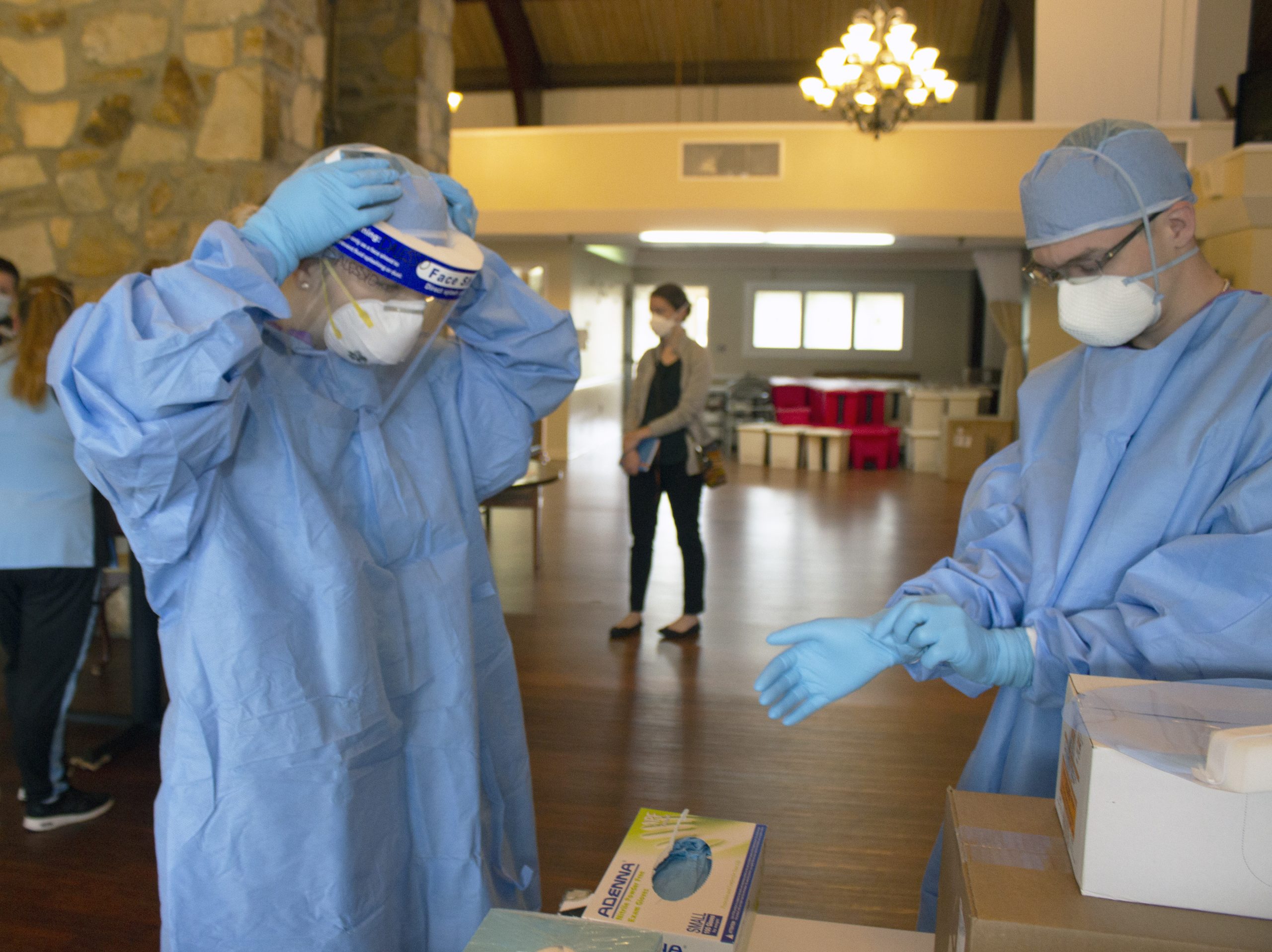
Medical asepsis is a term used to describe measures to prevent the spread of infection in health care agencies. Performing hand hygiene at appropriate times during patient care and applying gloves when there is potential risk for exposure to body fluids are examples of using medical asepsis. Additional precautions are implemented by health care team members when a patient has, or is suspected of having, an infectious disease. These additional precautions are called personal protective equipment (PPE) and are based on how an infection is transmitted, such as by contact, droplet, or airborne routes. Personal protective equipment (PPE) includes gowns, eyewear or goggles, face shields, gloves, and masks. PPE is used along with environmental controls, such as surface cleaning and disinfecting to prevent the transmission of infection.[13] See Figure 1.4[14] for an image of health care team members applying PPE. These precautions are further discussed in the “Aseptic Technique” chapter. For the purpose of this chapter, be sure to perform a general risk assessment before entering a patient’s room and apply the appropriate PPE as needed. This risk assessment includes the following:
- Is there signage posted on the patient’s door that contact, droplet, enhanced barrier, or airborne precautions are in place? If so, follow the instructions provided.
- Does this patient have a confirmed or suspected infection or communicable disease?
- Will your face, hands, skin, mucous membranes, or clothing be potentially exposed to blood or body fluids by spray, coughing, or sneezing?
Introducing Oneself
When initiating care with patients, it is essential to first provide privacy, and then introduce yourself and explain what will be occurring. Providing privacy means taking actions such as talking with the patient privately in a room with the door shut or privacy curtain drawn around the bed. A common framework used to communicate with patients is AIDET, a mnemonic for Acknowledge, Introduce, Duration, Explanation, and Thank You.[15]
- Acknowledge: Greet the patient by the name documented in their medical record. Make eye contact, smile, and acknowledge any family or friends in the room. Ask the patient their preferred way of being addressed (for example, "Mr. Doe," "Jonathon," or "Johnny") and their preferred pronouns (i.e., he/him, she/her, or they/them), as appropriate.
- Introduce: Introduce yourself by your name and role. For example, “I’m John Doe and I am a nursing student working with your nurse to take care of you today.”
- Duration: Estimate a timeline for how long it will take to complete the task you are doing. For example, “I am here to obtain your blood pressure, heart rate, and oxygen saturation levels. This should take about 5 minutes.”
- Explanation: Explain step by step what to expect next and answer questions. For example, “I will be putting this blood pressure cuff on your arm and inflating it. It will feel as if it is squeezing your arm for a few moments.”
- Thank You: At the end of the encounter, thank the patient and ask if anything is needed before you leave. In an acute or long-term care setting, ensure the call light is within reach and the patient knows how to use it. If family members are present, thank them for being there to support the patient as appropriate. For example, “Thank you for taking time to talk with me today. Is there anything I can get for you before I leave the room? Here is the call light (Place within reach). Press the red button if you would like to call the nurse.”
For more information about AIDET, visit AIDET Patient Communication.
Patient Identification
Use at least two patient identifiers before performing assessments, obtaining vital signs, or providing care.
Use two patient identifiers:
- Ask the patient to state their name and date of birth. If they have an armband, compare the information they are stating to the information on the armband and verify they match. See Figure 1.5[16] for an image of an armband.
- If the patient doesn’t have an armband, confirm the information they are stating to information provided in the chart.
- If the patient is unable to state their name and date of birth, scan their armband or ask another staff member or family member to identify them.
Confirm "two identifiers" with a second source:
- Scan the wristband.
- Compare the name and date of birth to the patient’s chart.
- Ask staff to verify the patient in a long-term care setting.
- Compare the picture on the medication administration record (MAR) to the patient.
- If present, ask a family member to confirm the patient’s name.

Cultural Safety
When initiating patient interaction, it is important to establish cultural safety. Cultural safety refers to the creation of safe spaces for patients to interact with health professionals without judgment or discrimination. See Figure 1.6[17] for an image representing cultural safety. Recognizing that you and all patients bring a cultural context to interactions in a health care setting is helpful when creating cultural safe spaces. If you discover you need more information about a patient’s cultural beliefs to tailor your care, use an open-ended question that allows the patient to share what they believe to be important. For example, you may ask, “I am interested in your cultural background as it relates to your health. Can you share with me what is important about your cultural background that will help me care for you?”[18]
For more information about caring for diverse patients, visit the "Diverse Patients" chapter in the Open RN Nursing Fundamentals textbook.

Adapting to Variations Across the Life Span
It is important to adapt your interactions with patients in accordance with their developmental stage. Developmentalists break the life span into nine stages[19]:
- Prenatal Development
- Infancy and Toddlerhood
- Early Childhood
- Middle Childhood
- Adolescence
- Early Adulthood
- Middle Adulthood
- Late Adulthood
- Death and Dying
A brief overview of the characteristics of each stage of human development is provided in Table 1.2. When caring for infants, toddlers, children, and adolescents, parents or guardians are an important source of information, and family dynamics should be included as part of the general survey assessment. When caring for older adults or those who are dying, other family members may be important to include in the general survey assessment. See Figure 1.7[20] for an image representing patients in various developmental stages of life.

Visit the Human Development Life Span e-book at LibreTexts to read additional information about human development across the life span.
Table 1.2 Variations Across the Life Span
| Stage of Development | Common Characteristics |
|---|---|
| Prenatal Development | Conception occurs, and development begins. All major structures of the body are forming, and the health of the mother is of primary concern. Understanding nutrition, teratogens (environmental factors that can lead to birth defects), and labor and delivery are primary concerns for the mother. |
| Infancy and Toddlerhood | The first year and a half to two years of life are ones of dramatic growth and change. A newborn with a keen sense of hearing but very poor vision is transformed into a walking, talking toddler within a relatively short period of time. Caregivers are also transformed from someone who manages feeding and sleep schedules to a constantly moving guide and safety inspector for a mobile, energetic child. |
| Early Childhood | Early childhood is also referred to as the preschool years, consisting of the years that follow toddlerhood and precede formal schooling. As a three- to five-year-old, the child is busy learning language, gaining a sense of self and greater independence, and beginning to learn the workings of the physical world. This knowledge does not come quickly however, and preschoolers may have initially interesting conceptions of size, time, space, and distance, such as fearing that they may go down the drain if they sit at the front of the bathtub. A toddler’s fierce determination to do something may give way to a four-year-old’s sense of guilt for doing something that brings the disapproval of others. |
| Middle Childhood | The ages of six through eleven comprise middle childhood, and much of what children experience at this age is connected to their involvement in the early grades of school. Their world becomes filled with learning and testing new academic skills, assessing one’s abilities and accomplishments, and making comparisons between self and others. Schools compare students and make these comparisons public through team sports, test scores, and other forms of recognition. Growth rates slow down, and children are able to refine their motor skills at this point in life. Children begin to learn about social relationships beyond the family through interaction with friends and fellow students. |
| Adolescence | The World Health Organization defines adolescence as a person between the age of 10 and 19. Adolescence is a period of dramatic physical change marked by an overall physical growth spurt and sexual maturation, known as puberty. It is also a time of cognitive change as the adolescent begins to think of new possibilities and to consider abstract concepts such as love, fear, and freedom. Adolescents have a sense of invincibility that puts them at greater risk of injury from high-risk behaviors such as car accidents, drug and alcohol abuse, or contracting sexually transmitted infections that can have lifelong consequences or result in death. |
| Early Adulthood | The twenties and thirties are often thought of as early adulthood. It is a time of physiological peak but also highest risk for involvement in violent crimes and substance abuse. It is a time of focusing on the future and putting a lot of energy into making choices that will help one earn the status of a full adult in the eyes of others. Love and work are primary concerns at this stage of life. |
| Middle Adulthood | The late thirties through the mid-sixties is referred to as middle adulthood. This is a period in which aging processes that began earlier become more noticeable but also a time when many people are at their peak of productivity in love and work. It can also be a time of becoming more realistic about possibilities in life previously considered and of recognizing the difference between what is possible and what is likely to be achieved in their lifetime. |
| Late Adulthood | This period of the life span has increased over the last 100 years. For nurses, patients in this period are referred to as “older adults.” The term “young old” is used to describe people between 65 and 79, and the term “old old” is used for those who are 80 and older. One of the primary differences between these groups is that the young old are very similar to midlife adults because they are still working, still relatively healthy, and still interested in being productive and active. The “old old” may remain productive, active, and independent, but risks of heart disease, lung disease, cancer, and cerebral vascular disease (i.e., strokes) increase substantially for this age group. Issues of housing, health care, and extending active life expectancy are only a few of the topics of concern for this age group. A better way to appreciate the diversity of people in late adulthood is to go beyond chronological age and examine whether a person is experiencing optimal aging (when they are in very good health for their age and continue to have an active, stimulating life), normal aging (when the changes in health are similar to most of those of the same age), or impaired aging (when more physical challenges and diseases occur compared to others of the same age). |
| Death and Dying | Death is the final stage of life. Dying with dignity allows an individual to make choices about treatment, say goodbyes, and take care of final arrangements. When caring for patients who are actively dying, nurses can advocate for care that allows that person to die with dignity according to their wishes. |
A body system review asks focused questions related to overall health status and body systems such as cardiac, respiratory, neurological, gastrointestinal, urinary, and musculoskeletal systems. See "Chapter Resources A" for a sample health history form that contains brief questions according to body systems. Nurses often incorporate review of system questions into the physical examination of each system. For example, while listening to bowel sounds in the abdomen, a nurse often inquires about the patient's bowel pattern. Additional focused assessment questions related to each body system are found in each assessment chapter of this book.
Information obtained during a health history interview is typically documented on agency-specific forms. See "Chapter Resources A" for a sample health history form used for documentation purposes. Additional information collected that is not included on the form should be documented in an associated progress note.
Functional health assessment collects data related to the patient’s functioning and their physical and mental capacity to participate in Activities of Daily Living (ADLs) and Instrumental Activities of Daily Living (IADLs). Activities of Daily Living (ADLs) are daily basic tasks that are fundamental to everyday functioning (e.g., hygiene, elimination, dressing, eating, ambulating/moving). See Figure 2.2[21] for an illustration of ADLs.

Instrumental Activities of Daily Living (IADL) are more complex daily tasks that allow patients to function independently such as managing finances, paying bills, purchasing and preparing meals, managing one’s household, taking medications, and facilitating transportation. See Figure 2.3[22] for an illustration of IADLs. Assessment of IADLs is particularly important to inquire about with young adults who have just moved into their first place, as well as with older patients with multiple medical conditions and/or disabilities.

Information obtained when assessing functional health provides the nurse a holistic view of a patient’s human response to illness and life conditions. It is helpful to use an assessment framework, such as Gordon’s Functional Health Patterns,[23] to organize interview questions according to evidence-based patterns of human responses. Using this framework provides the patient and their family members an opportunity to identify health-related concerns to the nurse that may require further in-depth assessment. It also verifies patient understanding of conditions so that misperceptions can be clarified. This framework includes the following categories:
- Nutritional-Metabolic: Food and fluid consumption relative to metabolic need
- Elimination: Excretion including bowel and bladder
- Activity-Exercise: Activity and exercise
- Sleep-Rest: Sleep and rest
- Cognitive-Perceptual: Cognition and perception
- Role-Relationship: Roles and relationships
- Sexuality-Reproductive: Sexuality and reproduction
- Coping-Stress Tolerance: Coping and effectiveness of managing stress
- Value-Belief: Values, beliefs, and goals that guide choices and decisions
- Self-Perception and Self-Concept: Self-concept and mood state[24]
- Health Perception-Health Management: A patient’s perception of their health and well-being and how it is managed. This is an umbrella category of all the categories above and underlies performing a health history.
The functional health section can be started by saying, “I would like to ask you some questions about factors that affect your ability to function in your day-to-day life. Feel free to share any health concerns that come to mind during this discussion.” Focused interview questions for each category are included in Table 2.8. Each category is further described below.
Nutrition
The nutritional category includes, but is not limited to, food and fluid intake, usual diet, financial ability to purchase food, time and knowledge to prepare meals, and appetite. This is also an opportune time to engage in health promotion discussions about healthy eating. Be aware of signs for malnutrition and obesity, especially if rapid and excessive weight loss or weight gain have occurred.
Life Span Considerations
When assessing nutritional status, the types of questions asked and the level of detail depend on the developmental age and health of the patient. Family members may also provide important information.
- Infants: Ask parents about using breast milk or formula, amount, frequency, supplements, problems, and introductions of new foods.
- Pregnant women: Include questions about the presence of nausea and vomiting and intake of folic acid, iron, omega-3 fatty acids, vitamin D, and calcium.
- Older adults or patients with disabling illnesses: Inquire about the ability to purchase and cook their food, decreased sense of taste, ability to chew or swallow foods, loss of appetite, and enough fiber and nutrients.[25]
Elimination
Elimination refers to the removal of waste products through the urine and stool. Health care professionals refer to urinating as voiding and stool elimination as having a bowel movement. Familiar terminology may need to be used with patients, such as “pee” and “poop.”
Constipation commonly occurs in hospitalized patients, so it is important to assess the date of their last bowel movement and monitor the frequency, color, and consistency of their stool.
Assess urine concentration, frequency, and odor, especially if concerned about urinary tract infection or incontinence. Findings that require further investigation include dysuria (pain or difficulty upon urination), blood in the stool, melena (black, tarry stool), constipation, diarrhea, or excessive laxative use.[26]
Life Span Considerations
When assessing elimination, the types of questions asked and the level of detail depends on the developmental age and health of the patient.
Toddlers: Ask parents or guardians about toilet training. Toilet training takes several months, occurs in several stages, and varies from child to child. It is influenced by culture and depends on physical and emotional readiness, but most children are toilet trained between 18 months and three years.
Older Adults: Constipation and incontinence are common symptoms associated with aging. Additional focused questions may be required to further assess these issues.[27]
Mobility, Activity, and Exercise
Mobility refers to a patient’s ability to move around (e.g., sit up, sit down, stand up, walk). Activity and exercise refer to informal and/or formal activity (e.g., walking, swimming, yoga, strength training). In addition to assessing the amount of exercise, it is also important to assess activity because some people may not engage in exercise but have an active lifestyle (e.g., walk to school or work in a physically demanding job).
Findings that require further investigation include insufficient aerobic exercise and identified risks for falls.[28]
Life Span Considerations
Mobility and activity depend on developmental age and a patient’s health and illness status. With infants, it is important to assess their ability to meet specific developmental milestones at each well-baby visit. Mobility can become problematic for patients who are ill or are aging and can result in self-care deficits. Thus, it is important to assess how a patient’s mobility is affecting their ability to perform ADLs and IADLs.[29]
Sleep and Rest
The sleep and rest category refers to a patient’s pattern of rest and sleep and any associated routines or sleeping medications used. Although it varies for different people and their life circumstances, obtaining eight hours of sleep every night is a general guideline. Findings that require further investigation include disruptive sleep patterns and reliance on sleeping pills or other sedative medications.[30]
Life Span Considerations
Older Adults: Disruption in sleep patterns can be especially troublesome for older adults. Assessing sleep patterns and routines will contribute to collaborative interventions for improved rest.[31]
Cognitive and Perceptual
The cognitive and perceptual category focuses on a person’s ability to collect information from the environment and use it in reasoning and other thought processes. This category includes the following:
- Adequacy of vision, hearing, taste, touch, feeling, and smell
- Any assistive devices used
- Pain level and pain management
- Cognitive functional abilities, such as orientation, memory, reasoning, judgment, and decision-making[32]
If a patient is experiencing pain, it is important to perform an in-depth assessment using the PQRSTU method described in the “Reason for Seeking Health Care” section of this chapter. It is also helpful to use evidence-based assessment tools when assessing pain, especially for patients who are unable to verbally describe the severity of their pain. See Figure 2.4[33] for an image of the Wong-Baker FACES tool that is commonly used in health care.
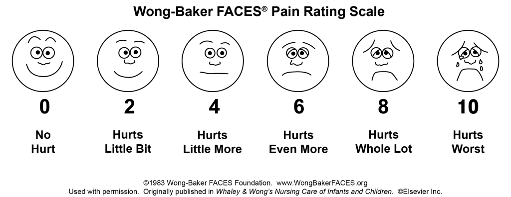
Life Span Considerations
Older Adults: Older adults are especially at risk for problems in the cognitive and perceptual category. Be alert for cues that suggest deficits are occurring that have not been previously diagnosed.
Roles - Relationships
Quality of life is greatly influenced by the roles and relationships established with family, friends, and the broader community. Roles often define our identity. For example, a patient may describe themselves as a “mother of an 8-year-old.” This category focuses on roles and relationships that may be influenced by health-related factors or may offer support during illness.[34] Findings that require further investigation include indications that a patient does not have any meaningful relationships or has “negative” or abusive relationships in their lives.
Life Span Considerations
Be sensitive to cues when assessing individuals with any of the following characteristics: isolation from family and friends during crisis, language barriers, loss of a significant person or pet, loss of job, significant home care needs, prolonged caregiving, history of abuse, history of substance abuse, or homelessness.[35]
Sexuality - Reproduction
Sexuality and sexual relations are an aspect of health that can be affected by illness, aging, and medication. This category includes a person’s gender identity and sexual orientation, as well as reproductive issues. It involves a combination of emotional connection, physical companionship (holding hands, hugging, kissing) and sexual activity that impact one’s feeling of health.[36]
The Joint Commission has defined terms to use when caring for diverse patients. Gender identity is a person’s basic sense of being male, female, or other gender.[37] Gender expression are characteristics in appearance, personality, and behavior that are culturally defined as masculine or feminine.[38] Sexual orientation is the preferred term used when referring to an individual’s physical and/or emotional attraction to the same and/or opposite gender.[39] LGBTQ is an acronym standing for the lesbian, gay, bisexual, transgender, and queer population. It is an umbrella term that generally refers to a group of people who are diverse in gender identity and sexual orientation. It is important to provide a safe environment to discuss health issues because the LGBTQ population experiences higher rates of smoking, alcohol use, substance abuse, HIV and other STD infections, anxiety, depression, suicidal ideation and attempts, and eating disorders as a result of stigma and marginalization.[40]
Life Span Considerations
Although sexuality is frequently portrayed in the media, individuals often consider these topics as private subjects. Use sensitivity when discussing these topics with different age groups across cultural beliefs while maintaining professional boundaries.
Advancing Effective Communication, Cultural Competence, and Patient- and Family-Centered Care for the Lesbian, Gay, Bisexual, and Transgender (LGBT) Community.
Coping-Stress Tolerance
Individuals experience stress that can lead to dysfunction if not managed in a healthy manner. Throughout life, healthy and unhealthy coping strategies are learned. Coping strategies are behaviors used to manage anxiety. Effective strategies control anxiety and lead to problem-solving but ineffective strategies can lead to abuse of food, tobacco, alcohol, or drugs.[41] Nurses teach and reinforce effective coping strategies.
Substance Use and Abuse
Alcohol, tobacco products, marijuana, and drugs are often used as ineffective coping strategies. It is important to use a nonjudgmental approach when assessing a patient’s use of substances, so they do not feel stigmatized. Substance abuse can affect people of all ages. Make a distinction between use and abuse as you assess frequency of use and patterns of behavior. Substance abuse often causes disruption in everyday function (e.g., loss of employment, deterioration of relationships, or precarious living circumstances) because of dependence on a substance. Action is needed if patients indicate that they have a problem with substance use or show signs of dependence, addiction, or binge drinking.[42]
Life Span Considerations
Some individuals are at increased risk for problems with coping strategies and stress management. Be sensitive to cues when assessing individuals with characteristics such as uncertainty in medical diagnosis or prognosis, financial problems, marital problems, poor job fit, or few close friends and family members.[43]
Value-Belief
This category includes values and beliefs that guide decisions about health care and can also provide strength and comfort to individuals. It is common for a person’s spirituality and values to be influenced by religious faith. A value is an accepted principle or standard of an individual or group. A belief is something accepted as true with a sense of certainty. Spirituality is a way of living that comes from a set of values and beliefs that are important to a person. The Joint Commission asks health care professionals to respect patients’ cultural and personal values, beliefs, and preferences and accommodate patients’ rights to religious and other spiritual services.[44] When performing an assessment, use open-ended questions to allow the patient to share values and beliefs they believe are important. For example, ask, “I am interested in your spiritual and religious beliefs and how they relate to your health. Can you share with me any spiritual beliefs or religious practices that are important to you during your stay?”
Self-Perception and Self-Concept
The focus of this category is on the subjective thoughts, feelings, and attitudes of a patient about themself. Self-concept refers to all the knowledge a person has about themself that makes up who they are (i.e., their identity). Self-esteem refers to a person’s self-evaluation of these items as being worthy or unworthy. Body image is a mental picture of one’s body related to appearance and function. It is best to assess these items toward the end of the interview because you will have already collected data that contributes to an understanding of the patient’s self-concept. Factors that influence a patient’s self-concept vary from person to person and include elements of life they value, such as talents, education, accomplishments, family, friends, career, financial status, spirituality, and religion.[45] The self-perception and self-concept category also focuses on feelings and mood states such as happiness, anxiety, hope, power, anger, fear, depression, and control.[46]
Life Span Considerations
Some individuals are at risk for problems with self-perception and self-concept. Be sensitive to cues when assessing individuals with characteristics such as uncertainty regarding a medical diagnosis or surgery, significant personal loss, history of abuse or neglect, loss of body part or function, or history of substance abuse.[47]
Violence and Trauma
There are many types of violence that a person may experience, including neglect or physical, emotional, mental, sexual, or financial abuse. You are legally mandated to report suspected cases of child abuse or neglect, as well as suspected cases of elder abuse. At any time, if you or the patient is in immediate danger, follow agency policy and procedure.
Trauma results from violence or other distressing events in a life. Collaborative intervention with the patient is required when violence and trauma are identified. People respond in different ways to trauma. It is important to use a trauma-informed approach when caring for patients who have experienced trauma. For example, a patient may respond to the traumatic situation in a way that seems unfitting (such as with laughter, ambivalence, or denial). This does not mean the patient is lying but can be a symptom of trauma. To reduce the effects of trauma, it is important to implement collaborative interventions to support patients who have experienced trauma.[48]
Loss of Body Part
A person can have negative feelings or perceptions about the characteristics, function, or limits of a body part as a result of a medical condition, surgery, trauma, or mental condition. Pay attention to cues, such as neglect of a body part or negative comments about a body part and use open-ended questions to obtain additional information.
Mental Health
Mental health is frequently underscreened and unaddressed in health care. The mental health of all patients should be assessed, even if they appear well or state they have no mental health concerns so that any changes in condition are quickly noticed and treatment implemented. Mental health includes emotional and psychological symptoms that can affect a patient's day-to-day ability to function. The World Health Organization (2014) defines mental health as “a state of well-being in which every individual realizes their own potential, can cope with normal stresses of life, can work productively and fruitfully, and is able to make a contribution to their community.”[49] Mental illness includes conditions diagnosed by a health care provider, such as depression, anxiety, addiction, schizophrenia, post-traumatic stress disorder, and others. Mental illness can disrupt everyday functioning and affect a person’s employment, education, and relationships.
It is helpful to begin this component of a mental health assessment with a statement such as, “Mental health is an important part of our lives, so I ask all patients about their mental health and any concerns or questions they may have.”[50] Be attentive of critical findings that require intervention. For example, if a patient talks about feeling hopeless or depressed, it is important to screen for suicidal thinking. Begin with an open-ended question, such as, “Have you ever felt like hurting yourself?” If the patient responds with a “Yes,” then progress with specific questions that assess the immediacy and the intensity of the feelings. For example, you may say, “Tell me more about that feeling. Have you been thinking about hurting yourself today? Have you put together a plan to hurt yourself?” When assessing for suicidal thinking, be aware that a patient most at risk is someone who has a specific plan about self-harm and can specify how and when they will do it. They are particularly at risk if planning self-harm within the next 48 hours. The age of the patient is not a factor in this determination of risk. If you believe the patient is at high risk, do not leave the patient alone. Collaborate with them regarding an immediate plan for emergency care.[51]
Health Perception-Health Management
Health perception-health management is an umbrella term encompassing all of the categories described above, as well as environmental health.
Environmental Health
Environmental health refers to the safety of a patient’s physical environment, also called a social determinant of health. Examples of environmental health include, but are not limited to, exposure to violence in the home or community; air pollution; and availability of grocery stores, health care providers, and public transportation. Findings that require further investigation include a patient living in unsafe environments.[52]
See Table 2.8 for sample focused questions for all categories related to functional health.[53]
Table 2.8 Focused Interview Questions for Functional Health Categories[54]
Begin this section by saying, "I would like to ask you some questions about factors that affect your ability to function in your day-to-day life. Feel free to share any health concerns that come to mind during this discussion.”
| Category | Focused Questions |
|---|---|
| Nutrition | Tell me about your diet.
What foods do you usually eat? What fluids do you usually drink every day? What have you eaten in the last 24 hours? Is this typical of your usual eating pattern? Tell me about your appetite. Have you had any changes in your appetite? Do you have any goals related to your nutrition? Do you have any financial concerns about purchasing food? Are you able to prepare the meals you want to eat? |
| Elimination | When was your last bowel movement?
Do you have any problems with constipation, diarrhea, or incontinence? Do you take laxatives or stool softeners? Do you have any problems urinating, such as frequent urination or burning on urination? Do you ever experience leaking or dribbling of urine? |
| Mobility, Activity, and Exercise | Tell me about your ability to move around.
Do you have any problems sitting up, standing up, or walking? Do you use any mobility aids (e.g., cane, walker, wheelchair)? Tell me about the activity and/or exercise in which you engage. What type? How frequent? For how long? |
| Sleep and Rest | Tell me about your sleep routine. How many hours of sleep do you usually get?
Do you feel rested when you awaken? Do you do anything to wind down before you go to bed (e.g., watch TV, read)? Do you take any sleeping medication? Do you take any naps during the day? |
| Cognitive and Perceptual | Are you having any pain?
Note: If present, use the PQRSTU method to further assess pain. Are you having any issues with seeing, hearing, smelling, tasting, or feeling things? Have you noticed any changes in memory or problems concentrating? Have you noticed any changes in the ability to make decisions? What is the easiest way for you to learn (e.g., written materials, explanations, or learning-by-doing)? |
| Roles and Relationships | Tell me about the most influential relationships in your life with family and friends.
How do these relationships influence your day-to-day life, health, and illness? Who are the people with whom you talk to when you require support or are struggling in your life? Do you have family or others dependent on you? Have you had any recent losses of someone important to you, a pet, or a job? Do you feel safe in your current relationship? |
| Sexuality-Reproduction | The expression of love and caring in a sexual relationship and creation of family are often important aspects in a person’s life. Do you have any concerns about your sexual health?
Tell me about the ways that you ensure your safety when engaging in intimate and sexual practices. |
| Coping-Stress | Tell me about the stress in your life.
Have you experienced a recent loss in your life that has impacted you? How do you cope with stress? |
| Values-Belief | I am interested in your spiritual and religious beliefs and how they relate to your health. Can you share with me any spiritual beliefs or religious practices that are important to you? |
| Self-Perception and Self-Concept |
Tell me what makes you who you are. How would you describe yourself? Have you noticed any changes in how you view your body or the things you can do? Are these a problem for you? Have you found yourself feeling sad, angry, fearful, or anxious? What helps you to feel better when this happens? Have you ever used any tobacco products (e.g., cigarettes, pipes, vaporizers, hookah)? If so, how much? How much alcohol do you drink every week? Have you used cannabis products? If so, how often do you use them? Have you ever used drugs or prescription drugs that were not prescribed for you? If so, what type? Have you ever felt you had a problem with any of these substances because they affected your daily life? If so, tell me more. Do you want to quit any of these substances? Many patients have experienced violence or trauma in their lives. Have you experienced any violence or trauma in your life? How has it affected you? Would you like to talk with someone about it?
|
| Health Perception - Health Management |
Tell me about how you take care of yourself and manage your home. Have you had any falls in the past six months? Do you have enough finances to pay your bills and purchase food, medications, and other needed items? Do you have any current or future concerns about being able to function independently? Tell me about where you live. Do you have any concerns about safety in your home or neighborhood? Tell me about any factors in your environment that may affect your health. Do you have any concerns about how your environment is affecting your health? |
Information obtained during a health history interview is typically documented on agency-specific forms. See "Chapter Resources A" for a sample health history form used for documentation purposes. Additional information collected that is not included on the form should be documented in an associated progress note.
Use the checklist below to review the steps for completion of "Obtaining a Health History."
Steps
Disclaimer: Always review and follow agency policy regarding this specific skill.
- Gather supplies: health history agency form.
- Knock, enter the room, greet the patient, and provide for privacy.
- Perform safety steps:
- Perform hand hygiene.
- Check the room for transmission-based precautions.
- Introduce yourself, your role, the purpose of your visit, and an estimate of the time it will take.
- Confirm patient ID using two patient identifiers (e.g., name and date of birth).
- Explain the process to the patient and ask if they have any questions.
- Be organized and systematic.
- Use appropriate listening and questioning skills.
- Listen and attend to patient cues.
- Ensure the patient's privacy and dignity.
- Assess ABCs.
- Address patient needs (pain, toileting, glasses/hearing aids) prior to starting. Note if the patient has signs of distress such as difficulty breathing or chest pain. If signs are present, defer the health history and obtain emergency assistance per agency policy.
- Complete a health history interview, including the following components per your instructor's instructions:
- Demographic and Biological Data
- Reason for Seeking Health Care
- Current and Past Medical History
- Family History
- Functional Health
- Review of Body Systems
- Ensure safety measures when leaving the room:
- CALL LIGHT: Within reach
- BED: Low and locked (in lowest position and brakes on)
- SIDE RAILS: Secured
- TABLE: Within reach
- ROOM: Risk-free for falls (scan room and clear any obstacles)
- Document the health history findings and report any concerns according to agency policy.
Learning Activities
(Answers to "Learning Activities" can be found in the "Answer Key" at the end of the book. Answers to interactive activity elements will be provided within the element as immediate feedback.)
Activities of daily living: Daily basic tasks fundamental to everyday functioning (e.g., hygiene, elimination, dressing, eating, ambulating/moving).
Belief: Something accepted as true with a sense of certainty.[55]
Body image: A mental picture of one’s body related to appearance and function.[56]
Care partners: Family and friends who are involved in helping to care for the patient.
Chief complaint: The reason a patient is seeking health care during a visit to a clinic or on admission to a health care facility.
Cultural safety: The creation of safe spaces for patients to interact with health professionals without judgment or discrimination.
Dysuria: Discomfort or pain on urinating.
Elimination: Refers to the removal of waste products through the urine and stool.
Functional health: The patient’s physical and mental capacity to participate in activities of daily living (ADLs) and instrumental activities of daily living (IADLs).
Gender expression: Characteristics in appearance, personality, and behavior, culturally defined as masculine or feminine.[57]
Gender identity: One’s basic sense of being male, female, or other gender.[58]
Health history: The process of using directed interview questions to obtain symptoms and perceptions about a patient’s illness or life condition. The purpose of obtaining a health history is to gather subjective data from the patient and/or the patient’s family so that the health care team and the patient can collaboratively create a plan that will promote health, address acute health problems, and minimize chronic health conditions.
Instrumental activities of daily living: Complex daily tasks that allow patients to function independently such as managing finances, paying bills, purchasing and preparing meals, managing one’s household, taking medications, and facilitating transportation.
LGBTQ: An acronym standing for lesbian, gay, bisexual, transgender, and queer is an umbrella term that generally refers to a group of people who are diverse with regard to their gender identity and sexual orientation. There are expanded versions of this acronym.[59]
Main health care needs: Term used to classify what needs the patient feels are most important to address after admission to a health care agency.
Medication reconciliation: A comparison of a list of current medications with a previous list and is completed at every hospitalization and clinic visit.
Melena: Dark, tarry-looking stool due to the presence of digested blood.
Mental health: A state of well-being in which every individual realizes their own potential, can cope with normal stresses of life, can work productively and fruitfully, and is able to make a contribution to their community.[60]
Mobility: A patient’s ability to move around (e.g., sit up, sit down, stand up, walk).
Nursing: Nursing integrates the art and science of caring and focuses on the protection, promotion, and optimization of health and human functioning; prevention of illness and injury; facilitation of healing; and alleviation of suffering through compassionate presence. Nursing is the diagnosis and treatment of human responses and advocacy in the care of individuals, families, groups, communities, and populations in recognition of the connection of all humanity.[61]
Objective data: Information observed through your sense of hearing, sight, smell, and touch while assessing the patient.
Primary source of data: Information obtained directly from the patient.
Secondary source of data: Information from the patient's chart, family members, or other health care team members.
Self-concept: Knowledge a person has about themselves that makes up who they are (i.e., their identity).
Self-esteem: A person’s self-evaluation of their self-concept as being worthy or unworthy.
Sexual orientation: The preferred term used when referring to an individual's physical and/or emotional attraction to the same and/or opposite gender.[62]
Sign: Objective data found by the nurse or health care provider when assessing a patient.
Spirituality: A way of living that comes from a set of meanings, values, and beliefs that are important to a person.[63]
Subjective data: Information obtained from the patient and/or family members that offers important cues from their perspectives.
Symptom: Subjective data that the patient reports, such as “I feel dizzy.”
Value: An accepted principle or standard of an individual or group.[64]
Voiding: Medical terminology used for urinating.
Activities of daily living: Daily basic tasks fundamental to everyday functioning (e.g., hygiene, elimination, dressing, eating, ambulating/moving).
Belief: Something accepted as true with a sense of certainty.[65]
Body image: A mental picture of one’s body related to appearance and function.[66]
Care partners: Family and friends who are involved in helping to care for the patient.
Chief complaint: The reason a patient is seeking health care during a visit to a clinic or on admission to a health care facility.
Cultural safety: The creation of safe spaces for patients to interact with health professionals without judgment or discrimination.
Dysuria: Discomfort or pain on urinating.
Elimination: Refers to the removal of waste products through the urine and stool.
Functional health: The patient’s physical and mental capacity to participate in activities of daily living (ADLs) and instrumental activities of daily living (IADLs).
Gender expression: Characteristics in appearance, personality, and behavior, culturally defined as masculine or feminine.[67]
Gender identity: One’s basic sense of being male, female, or other gender.[68]
Health history: The process of using directed interview questions to obtain symptoms and perceptions about a patient’s illness or life condition. The purpose of obtaining a health history is to gather subjective data from the patient and/or the patient’s family so that the health care team and the patient can collaboratively create a plan that will promote health, address acute health problems, and minimize chronic health conditions.
Instrumental activities of daily living: Complex daily tasks that allow patients to function independently such as managing finances, paying bills, purchasing and preparing meals, managing one’s household, taking medications, and facilitating transportation.
LGBTQ: An acronym standing for lesbian, gay, bisexual, transgender, and queer is an umbrella term that generally refers to a group of people who are diverse with regard to their gender identity and sexual orientation. There are expanded versions of this acronym.[69]
Main health care needs: Term used to classify what needs the patient feels are most important to address after admission to a health care agency.
Medication reconciliation: A comparison of a list of current medications with a previous list and is completed at every hospitalization and clinic visit.
Melena: Dark, tarry-looking stool due to the presence of digested blood.
Mental health: A state of well-being in which every individual realizes their own potential, can cope with normal stresses of life, can work productively and fruitfully, and is able to make a contribution to their community.[70]
Mobility: A patient’s ability to move around (e.g., sit up, sit down, stand up, walk).
Nursing: Nursing integrates the art and science of caring and focuses on the protection, promotion, and optimization of health and human functioning; prevention of illness and injury; facilitation of healing; and alleviation of suffering through compassionate presence. Nursing is the diagnosis and treatment of human responses and advocacy in the care of individuals, families, groups, communities, and populations in recognition of the connection of all humanity.[71]
Objective data: Information observed through your sense of hearing, sight, smell, and touch while assessing the patient.
Primary source of data: Information obtained directly from the patient.
Secondary source of data: Information from the patient's chart, family members, or other health care team members.
Self-concept: Knowledge a person has about themselves that makes up who they are (i.e., their identity).
Self-esteem: A person’s self-evaluation of their self-concept as being worthy or unworthy.
Sexual orientation: The preferred term used when referring to an individual's physical and/or emotional attraction to the same and/or opposite gender.[72]
Sign: Objective data found by the nurse or health care provider when assessing a patient.
Spirituality: A way of living that comes from a set of meanings, values, and beliefs that are important to a person.[73]
Subjective data: Information obtained from the patient and/or family members that offers important cues from their perspectives.
Symptom: Subjective data that the patient reports, such as “I feel dizzy.”
Value: An accepted principle or standard of an individual or group.[74]
Voiding: Medical terminology used for urinating.
View a Sample Health History Form.
View a Sample Health History Form.
The accurate measurement of blood pressure is important for ensuring patient safety and optimizing body system function. Blood pressure measurements are used by health care providers to make important decisions about a patient’s care. Blood pressure measurements help providers make decisions about whether a patient needs fluids or prescription medications. It is crucial to follow the proper steps to obtain a patient’s blood pressure to ensure the care team has accurate data to help make health care decisions and determine a plan of care.

